Review
. 2025 Jun 26;12(7):245.
doi: 10.3390/jcdd12070245.

The Anatomy of the Atrioventricular Node

Affiliations
Review

The Anatomy of the Atrioventricular Node

Robert H Anderson et al. J Cardiovasc Dev Dis. .

Abstract

The anatomical arrangement of the atrioventricular node has been likened to a riddle wrapped up in an enigma. There are several reasons for this alleged mystery, not least the marked variability in structure between different species. Lack of detailed knowledge of the location of the node relative to the atrial and ventricular septal structures has also contributed to previous misunderstandings. Recent studies comparing the findings of gross dissection with virtual dissection of living datasets, combined with access to a large number of serially sectioned human and animal hearts, have served to provide the evidence to solve the riddle. We summarise these findings in this review. We explain how the node is located within the atrial walls of the inferior pyramidal space. It becomes the non-branching component of the atrioventricular conduction axis as the axis extends through the plane of atrioventricular insulation to enter the infero-septal recess of the left ventricular outflow tract. The node itself is formed by contributions from the tricuspid and mitral vestibules, with extensive additional inputs from the base of the atrial septum. We show how knowledge of development enhances the appreciation of the arrangements and offers an explanation as to why, on occasion, there can be persisting nodoventricular connections. We discuss the findings relative to the circuits producing atrioventricular re-entry tachycardia. We conclude by emphasising the significance of the variation of the anatomical arrangements within different mammalian species.

Keywords: compact node; fast pathway; inferior extensions; inferior pyramidal space; infero-septal recess; slow pathway.


Conflict of interest statement

The authors declare no conflicts of interest.

Figures

Figure 2
Figure 2
(A) Panel A shows the drawing made by Walter Koch [21], in which he shows the boundaries of the triangle we now describe in his name as providing the landmarks to the atrioventricular node (compare with Figure 1). (B) Panel B is a reproduction of one of the drawings made by Sunao Tawara to show the atrioventricular node, which he described as the Knoten [19], as the atrial origin of an atrioventricular conduction axis. We have reorientated the figure as it would be seen in the short axis of the ventricular mass, viewed from the apex of the heart looking towards the base. (C) Panel C is a section taken in the short axis of an adult human heart. It shows the accuracy of the arrangement as depicted by Tawara. The section also serves to demonstrate the answer to a potential paradox. As viewed in the drawing provided by Koch, the node is seen to be located centrally within the cardiac mass. The short axis shown in panel C confirms that to be the case. At the same time, however, the node is directly related to the epicardial tissues of the atrioventricular grooves. It is positioned within the atrial walls at the apex of the inferior pyramidal space.
Figure 1
Figure 1
(A) Panel A shows the septal surfaces of the right atrium and ventricle of an adult human heart having removed the parietal walls of the chambers. The specimen is transilluminated from the left side to show the locations of the oval fossa and the membranous septum. The dashed lines show the locations of the tendon of Todaro and the hinge of the septal leaflet of the tricuspid valve. As we will describe, these structures form the boundaries of the triangle established by Koch as providing the landmarks to the atrioventricular node (see Figure 2A). The double headed arrow shows the location of the septal isthmus, an area that will become significant when we discuss the substrates for atrioventricular re-entry tachycardia. (B) In panel B, careful dissection has revealed the location of the atrioventricular node within the walls of the triangle and shown its continuation as the right bundle branch. When using gross dissection, however, there is no certainty that the pathway demonstrated is the solitary pathway for atrioventricular conduction.
Figure 3
Figure 3
(A) In panel A, the potential septal surfaces of the right atrium and ventricle at the level of the atrioventricular junction have been revealed by opening and spreading the junction. (B) The dissection shown in panel B has removed all the walls of the right atrium that, on their posterior aspect, are bordered by the fibro-adipose tissues of the extramural areas, themselves enclosed within the epicardial coverings of the heart. The dissection shows how the atrial septum is formed primarily by the floor of the oval fossa, along with its anterior buttress, which binds the floor of the fossa, derived from the primary atrial septum, to the insulating tissues of the atrioventricular junction. The remaining rims of the fossa are infoldings of the atrial walls. The so-called sinus septum, also known as the Eustachian ridge, is similarly an infolding between the walls of the inferior caval vein and the coronary sinus. Removal of the tricuspid vestibule shows how it forms one of the boundaries of the inferior pyramidal space, which is filled with the fibro-adipose tissues of the inferior atrioventricular groove.
Figure 4
Figure 4
(A) The dissection of an infant heart offers further insights to the boundaries of the inferior pyramidal space. To produce the image shown in panel A, a cut had been made to liberate the right atrial wall from its posterior neighbours. (B) The cut was directed obliquely into the aortic root, producing the image shown in Panel B, which shows the posterior parts as seen from the right side. The cut has revealed the extent of the fibro-adipose tissues filling the inferior pyramidal space, which is limited cranially by the buttress of the atrial septum. The cut through the aortic root has also revealed the location of the infero-septal recess of the left ventricular outflow tract. Note that the walls of the coronary sinus are surrounded by the fibro-adipose tissues of the inferior pyramidal space. (C) Panel C shows the left side of the segment removed by the cut that liberated the right atrial structures from the posterior neighbours. The atrioventricular node is contained within the tissues shown behind the wall of the triangle of Koch as shown in panel A. As we will describe, it penetrates at the level of the membranous septum to enter the infero-septal recess as the non-branching atrioventricular bundle.
Figure 5
Figure 5
The section is from the same series as shown as panel C of Figure 2. This section shows how the right atrial vestibule is separated by the fibro-adipose tissues of the inferior pyramidal space from the parietal wall of the left ventricle, which forms the ventricular boundary of the space. Compare with Figure 6B.
Figure 6
Figure 6
(A) Panel A shows the anatomist’s view of the atrioventricular junctions, having cut back the atrial walls to the level of the vestibules. The right atrial wall of Koch’s triangle can be judged to overlie the fibro-adipose tissues of the inferior pyramidal space, with the base of the pyramid formed by the cardiac crux, and its apex by the atrial septal buttress. (B) Panel B shows the view that can now be obtained by segmentation of living computed tomographic datasets. The image has been orientated so as to match the view shown in panel A. The wall of Koch’s triangle and the roof of the infero-septal recess have been segmented. As can be seen in panel B, the base of the pyramid is the inferior surface of the heart.
Figure 7
Figure 7
(A) Panel A shows a cross section through the walls of the right atrium from a heart obtained from a 6-month-old neonate, with the image orientated in “four chamber” fashion. Even at this low magnification it is possible to recognise the cardiomyocytes making up the atrioventricular node and the site of the sinus node, although the cells of the sinus node are not seen. The remaining walls are made up of aggregated working cardiomyocytes, with an obvious parallel alignment in the right atrial wall making up part of the superior rim of the oval fossa. (B) Panel B shows a dissection of the septal surface of the right atrium, having removed the endocardium to show the “grain” produced by the aggregation of the cardiomyocytes. There are obvious parallel alignments in the pathways leading into the triangle of Koch.
Figure 8
Figure 8
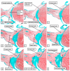
(AI) The panels are selected sections from the serially prepared dataset obtained from the heart from the neonate of 6 months shown in lower magnification in Figure 7A. To make this figure, the images are orientated in an attitudinally appropriate fashion, with the right side to the top. Panel A shows a section at the base of the pyramid of Koch, with subsequent sections moving cranially to panel I, which is close to the apex of the pyramid.
Figure 9
Figure 9
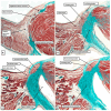
(AD) The histological sections, again shown in a serial fashion extending from the base of the pyramid of Koch (panel A) to its apex (Panel D), are taken from an adult heart. The sections are again orientated in an attitudinally appropriate fashion, with the right-sided chambers to the top of the image.
Figure 10
Figure 10
The panels show the key stages in remodelling of the embryonic interventricular communication relative to the formation of the atrioventricular node. The panel seen to the left hand shows a view, as seen from the apex looking towards the base, of the ring of specialised cardiomyocytes that surround the embryonic interventricular foramen prior to expansion of the atrioventricular canal. The panel seen to the right hand shows the arrangement subsequent to expansion to form the right atrioventricular junction, and as the aortic root begins to translocate to achieve its eventual position within the left ventricle. The reconstructions are made from the interactive pdf files available from the publication of Hikspoors and colleagues [33].
Figure 11
Figure 11
The images show histological sections stained with hematoxylin and eosin, in four-chamber orientation, revealing the arrangement of the primary ring at its inferior transition from the ventricular to the atrial components of the developing heart. (A) Panel A shows the arrangement at Carnegie stage 17, equal to around 6 weeks of gestation. At this stage, the vestibular spine and mesenchymal cap have still to muscularise. (B) Panel B shows the situation at the end of the embryonic period of development. The spine and cap have muscularised and provide the septal inputs to the compact node, which is derived from the primary ring. The ring extends between the horns of the inferior atrioventricular cushion as the ring itself transitions to become the compact atrioventricular node. At this stage, the insulation between the base of the node and the crest of the muscular septum has yet to be formed.
Figure 12
Figure 12
The panels show the major changes required to transform the arrangement at the end of the embryonic period of development (panel A) to the definitive arrangement (panel B). (A) Panel A is a reconstruction of an embryonic heart at 8 weeks of development. There has been no expansion of the atrioventricular junctions to produce the inferior pyramidal space, and the aortic root has yet to be “wedged” between the superior parts of the right and left atrioventricular junctions. Note how the primary ring emerges from beneath the inferior atrioventricular cushion to enter the tricuspid vestibule (See also Figure 11B). Both vestibules, however, are derived from atrioventricular canal myocardium, which is slowly conducting. (B) Panel B shows a virtual dissection of a computed tomographic dataset from a living patient. It is the expansion of the junctions that produces the triangle of Koch and the inferior pyramidal space. Note how the virtual basal ring of the aortic root has become wedged between the superior components of the junctions.
Figure 13
Figure 13
The images are taken from a human heart scanned using HiP-CT technology at the European Synchrotron Radiation Facility, made available at 19.89 um, in this case, of the heart of the body donor s-20-29 [3]. (A) Panel A is a two-dimensional image, showing the transition from the atrioventricular node at the apex of the inferior pyramidal space, through the non-branching bundle located within the infero-septal recess, and to the continuation as the right bundle branch. (B) Panel B then shows how it is possible to reconstruct these features within the heart itself, showing the relationship of the ventricular components of the axis to the virtual basal ring of the aortic root.
Figure 14
Figure 14
The images are taken from a human heart scanned using HiP-CT at the European Synchrotron Radiation Facility and made available at 6.51 um from the heart of the body donor S20-29. (A) Panel A shows the transition between the compact node and the rightward extension into the right atrial vestibule, together with the merge-point between the rightward extension and working myocardium. (B) Panel B shows the input to the compact node from the atrial septum, in other words the fast-pathway, with minor infiltration by fat separating it from the right atrial vestibule. (C) Panel C shows the much smaller leftward extension from the compact node and the merge-point with the working myocardium of the left atrial vestibule.
Figure 15
Figure 15
The images show histological sections prepared from three neonatal hearts from patients with Ebstein’s malformation [47]. All sections are orientated in comparable fashion to Figure 7 and Figure 8. (A) Panel A shows dispersion of the nodal cardiomyocytes throughout the fibrous atrioventricular insulating tissues, with a strand producing a nodoventricular communication. (B) In panel B, there is less dispersion but a more obvious nodoventricular pathway. (C) In panel C, there is still less dispersion, but the nodoventricular pathway is insulated as it extends into the crest of the muscular ventricular septum.
Figure 16
Figure 16
(A) Panel A shows the potential circuits for atrioventricular nodal re-entry superimposed on a reconstruction from a living computed tomographic dataset. (B) The ideal site for placing an ablative lesion is shown in panel B, where the lesion placed at the septal isthmus terminated the re-entry circuit and cured the tachycardia. Histological examination revealed that the ablation had involved working atrial cardiomyocytes [50].
Figure 17
Figure 17
(AC) The serial histological sections, prepared in a fashion comparable to the arrangements shown in Figure 7, Figure 8 and Figure 15, reveal that the arrangement of the conduction axis in the murine heart, unlike the situation in the canine and rabbit hearts, is comparable to the arrangement as found in the human heart.

References

    1. Anderson R.H., Sanchez-Quintana D., Mori S., Cabrera J.A., Back Sternick E. Re-evaluation of the structure of the atrioventricular node and its connections with the atrium. EP Eur. 2020;22:821–830. doi: 10.1093/europace/euaa031. - DOI - PubMed
    1. Katritsis D.G. The Human Atrioventricular Node: Oedipus and the Riddle of the Sphinx. Arrhythmia Electrophysiol. Rev. 2020;9:52–53. doi: 10.15420/aer.2020.30. - DOI - PMC - PubMed
    1. Brunet J., Cook A.C., Walsh C.L., Cranley J., Tafforeau P., Engel K., Arthurs O., Berruyer C., Burke O’Leary E., Bellier A., et al. Multidimensional analysis of the adult human heart in health and disease using hierarchical phase-contrast tomography. Radiology. 2024;312:e232731. doi: 10.1148/radiol.232731. - DOI - PMC - PubMed
    1. Anderson R.H., Mori S., Spicer D.E., Sanchez-Quintana D., Jensen B. The anatomy, development, and evolution of the atrioventricular conduction axis. J. Cardiovasc. Dev. Dis. 2018;5:44. doi: 10.3390/jcdd5030044. - DOI - PMC - PubMed
    1. Tretter J.T., Spicer D.E., Sánchez-Quintana D., Back Sternick E., Farré J., Anderson R.H. Miniseries 1—Part III:‘Behind the scenes’ in the triangle of Koch. EP Eur. 2022;24:455–463. doi: 10.1093/europace/euab285. - DOI - PubMed

LinkOut - more resources