. 2025 Jun 26;13(7):538.
doi: 10.3390/toxics13070538.

NOX2/NLRP3-Inflammasome-Dependent Microglia Activation Promotes As(III)-Induced Learning and Memory Impairments in Developmental Rats

Affiliations

NOX2/NLRP3-Inflammasome-Dependent Microglia Activation Promotes As(III)-Induced Learning and Memory Impairments in Developmental Rats

Linlin Zhang et al. Toxics. .

Abstract

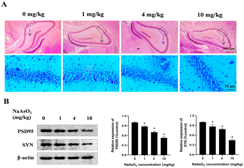
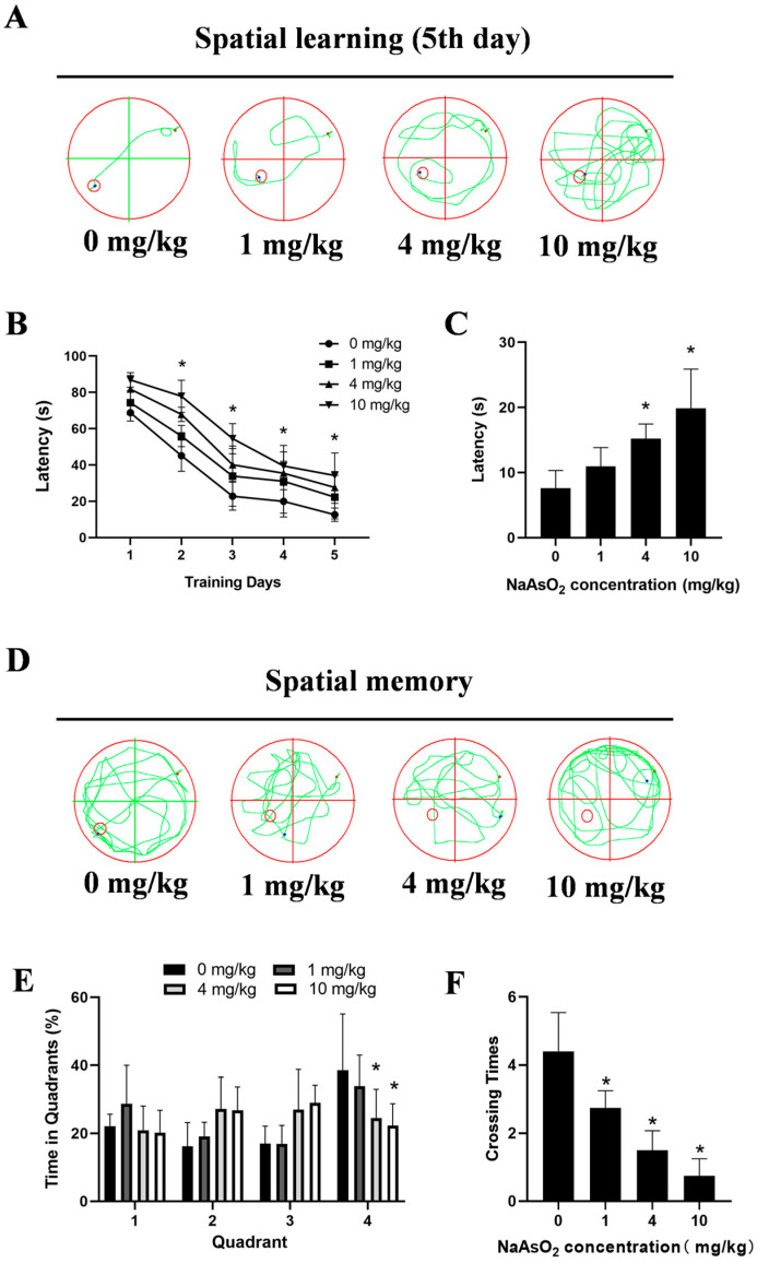
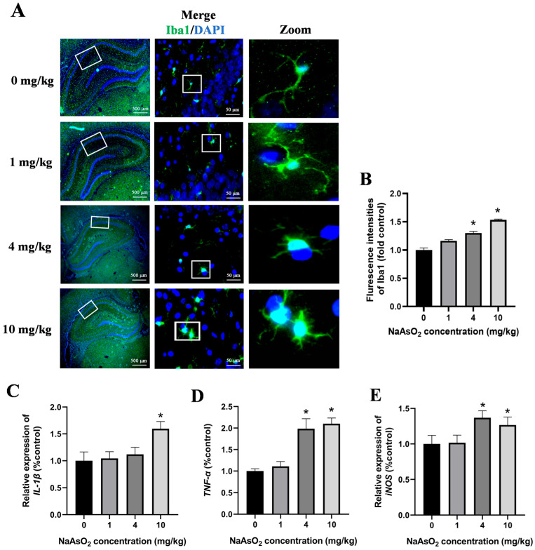
Inorganic arsenic [As(III) and As(V)] is a pervasive environmental contaminant in groundwater systems, early-life exposure to which is associated with an impaired cognitive ability and an increased risk of neurobehavioral disorders. Although the effect of As(III) on the neurons is well studied, the involvement of the microglia remains unclear. In this study, the effects of sodium arsenite (NaAsO2) on microglial activation and the underlying NLRP3 inflammasome mechanism were determined. Pregnant rats were gavaged with NaAsO2 (0, 1, 4, and 10 mg/kg body weight), which dissociates in aqueous solutions into bioactive arsenite species [As(OH)3], from gestational day 1 (GD1) to postnatal day 21 (PND21). The results showed that As(III) induces learning and memory impairments and microglial activation in the hippocampus of offspring rats (PND21). Increased expression of NLRP3, the activation of caspase-1, and the production of interleukin-1β were observed in both the hippocampus of As(III)-exposed offspring rats and As(III)-exposed microglial BV2 cells under culture conditions. Interestingly, blocking the NLRP3 inflammasome using MCC950 mitigated its activation. Furthermore, inhibition of NADPH oxidase 2 (NOX2) using apocynin or specific siRNA significantly reduced As(III)-induced microglial NLRP3 inflammasome activation. In addition, inactivation of the microglial NLRP3 inflammasome or NOX2 markedly rescued As(III)-induced neurotoxicity in the hippocampal HT22 cells. Taken together, this study reveals that NOX2/NLRP3-inflammasome-dependent microglial activation promotes As(III)-induced learning and memory impairments in developmental rats.

Keywords: NLRP3 inflammasome; NOX2; inorganic arsenic; learning and memory; microglia.


Conflict of interest statement

The authors declare no conflicts of interest.

Figures

Figure 1
Figure 1
Gestational/lactational NaAsO2 exposure induces hippocampal tissue damage in offspring rats. SD rats were gavaged with NaAsO2 (0, 1, 4, and 10 mg/kg body weight) from gestational day 1 to postnatal day 21 (PND21). Offspring was sacrificed on PND21. (A) Pathological changes in the hippocampus were observed through HE staining (scale bar = 500 μm, 4× magnification; scale bar = 50 μm, 20× magnification; arrow, the dashed box area enlarged below). (B) PSD95 and SYN in the hippocampus were detected using a Western blot analysis. Full Western blot images can be found in Figure S1. The data are expressed as mean ± standard error (SEM). n = 3–12 in each group; * p < 0.05 vs. control.
Figure 2
Figure 2
Gestational/lactational NaAsO2 exposure induces learning–memory deficits in offspring rats. SD rats were gavaged with NaAsO2 (0, 1, 4, and 10 mg/kg body weight) from gestational day 1 to postnatal day 21 (PND 21). PND21 rats were subjected to the Morris water maze test. (A) Day 5 learning path panel. Small red circle, escape platform location; small red dot, rat entry point; small blue dot, rat exit point. (B) Escape latency in the first 5 days. (C) Escape latency in the probe test. (D) Probe trial path panel. Small red circle, escape platform location; small red dot, rat entry point; small blue dot, rat exit point. (E) Quadrant times in the probe test. (F) Crossings to the original platform location. Data are expressed as the mean ± SEM. n = 10–12 in each group; * p < 0.05 vs. control.
Figure 3
Figure 3
Gestational/lactational NaAsO2 exposure causes microglial activation in the hippocampus of offspring rats. (A) Immunofluorescence staining using the anti-Iba-1 antibody was performed, and representative images are shown (scale bar = 500 μm, 4× magnification; scale bar = 50 μm, 20× magnification; white box, the area magnified in the right panel.). (B) The density of green fluorescence of Iba1 was quantified. The (C) IL-1β, (D) TNF-α, and (E) iNOS gene expression in the hippocampus was measured through real-time PCR. Data are expressed as the mean ± SEM. n = 3–6 in each group; * p < 0.05 vs. control.
Figure 4
Figure 4
The NLRP3 inflammasome contributes to NaAsO2-induced activation of the microglia. (A) NLRP3, pro-caspase-1, pro-IL-1β, ASC, caspase-1, and mature IL-1β in the hippocampus of NaAsO2-exposed offspring rats were detected through a Western blot analysis, and (B) the density of the blots was quantified. Full Western blot images can be found in Figure S2. (C) BV2 microglial cells were treated with 0, 0.5, and 2 μM of NaAsO2. After 24 h, the expression of NLRP3, pro-caspase-1, pro-IL-1β, ASC, caspase-1, and mature IL-1β was detected through a Western blot analysis, and (D) the density of the blots was quantified. Full Western blot images can be found in Figure S3. (E) BV2 microglial cells were treated with 2 μM of NaAsO2 with or without MCC950 (an inhibitor of the NLRP3 inflammasome). After 24 h, the gene expression of (E) IL-1β, (F) TNF-α, and (G) iNOS was measured through real-time PCR. Data are expressed as the mean ± SEM. n = 3–6 in each group; * p < 0.05 vs. control, # p < 0.05 vs. the As group.
Figure 5
Figure 5
NOX2 is key to NLRP3 inflammasome activation in NaAsO2-exposed microglia. The NOX2 gene expression in the hippocampus (A) and BV2 microglial cells (B) was measured through real-time PCR. (C) BV2 microglial cells were treated with 2 μM of NaAsO2 with or without APO pretreatment (a NOX2 inhibitor). After 24 h, the expression of NLRP3, pro-caspase-1, pro-IL-1β, ASC, caspase-1, and mature IL-1β was detected through a Western blot analysis, and (D) the density of the blots was quantified. Full Western blot images can be found in Figure S4. (E) BV2 microglial cells were treated with 2 μM of NaAsO2 with or without siNOX2. After 24 h, the expression of NLRP3, pro-caspase-1, pro-IL-1β, ASC, caspase-1, and mature IL-1β was detected through a Western blot analysis, and (F) the density of the blots was quantified. Full Western blot images can be found in Figure S5. Data are expressed as the mean ± SEM. n = 3–6 in each group; * p < 0.05 vs. Control, # p < 0.05 vs. the As group.
Figure 6
Figure 6
Inhibition of the microglial NLRP3 inflammasome or NOX2 mitigates NaAsO2-induced impairments in hippocampal HT22 cells. BV2 microglial cells were treated with 2 μM of NaAsO2 with or without MCC950 or APO pretreatment. After 24 h, the conditioned medium (CM) was collected and then added to the hippocampal HT22 cells. The PSD95 and SYN levels in the HT22 cells, important mediators of learning and memory, were measured using a Western blot analysis after 48 h of CM treatment. Full Western blot images can be found in Figure S6. Data are expressed as the mean ±S EM. n = 3–4 in each group; * p < 0.05 vs. control, # p < 0.05 vs. the As group.

References

    1. Podgorski J., Berg M. Global threat of arsenic in groundwater. Science. 2020;368:845–850. doi: 10.1126/science.aba1510. - DOI - PubMed
    1. Rahaman M.S., Rahman M.M., Mise N., Sikder M.T., Ichihara G., Uddin M.K., Kurasaki M., Ichihara S. Environmental arsenic exposure and its contribution to human diseases, toxicity mechanism and management. Environ. Pollut. 2021;289:117940. doi: 10.1016/j.envpol.2021.117940. - DOI - PubMed
    1. Martinez V.D., Lam W.L. Health Effects associated with pre- and perinatal exposure to arsenic. Front. Genet. 2021;12:664717. doi: 10.3389/fgene.2021.664717. - DOI - PMC - PubMed
    1. Sanders A.P., Desrosiers T.A., Warren J.L., Herring A.H., Enright D., Olshan A.F., Meyer R.E., Fry R.C. Association between arsenic, cadmium, manganese, and lead levels in private wells and birth defects prevalence in North Carolina: A semi-ecologic study. BMC Public Health. 2014;14:955. doi: 10.1186/1471-2458-14-955. - DOI - PMC - PubMed
    1. Farzan S.F., Karagas M.R., Chen Y. In utero and early life arsenic exposure in relation to long-term health and disease. Toxicol. Appl. Pharmacol. 2013;272:384–390. doi: 10.1016/j.taap.2013.06.030. - DOI - PMC - PubMed

LinkOut - more resources