. 2025 Jun 20;12(7):602.
doi: 10.3390/vetsci12070602.

The Giant Panda Transferrin Receptor Facilitates Feline Parvovirus Infection to Drive Cross-Species Transmission

Affiliations

The Giant Panda Transferrin Receptor Facilitates Feline Parvovirus Infection to Drive Cross-Species Transmission

Qigui Yan et al. Vet Sci. .

Abstract

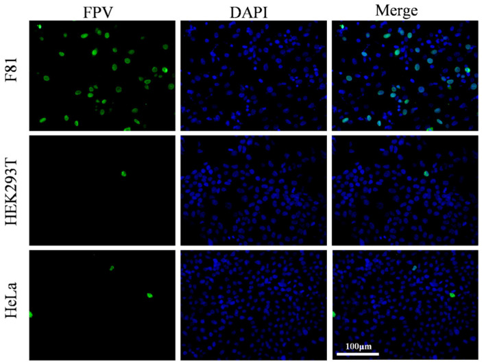
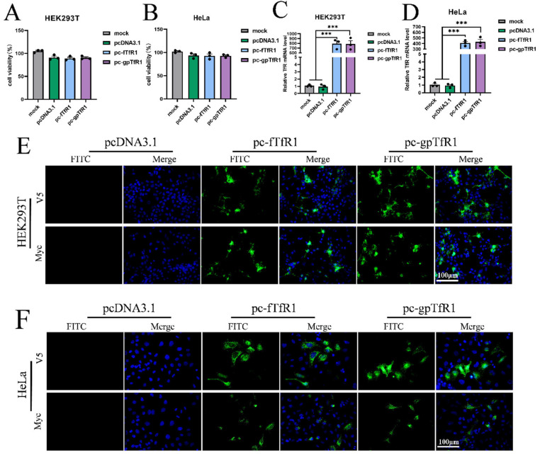
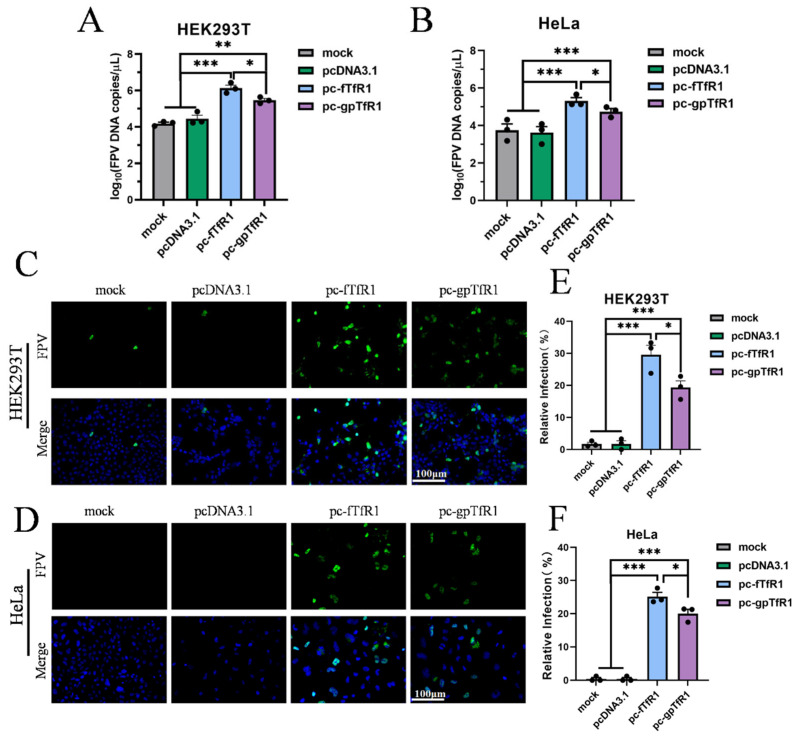
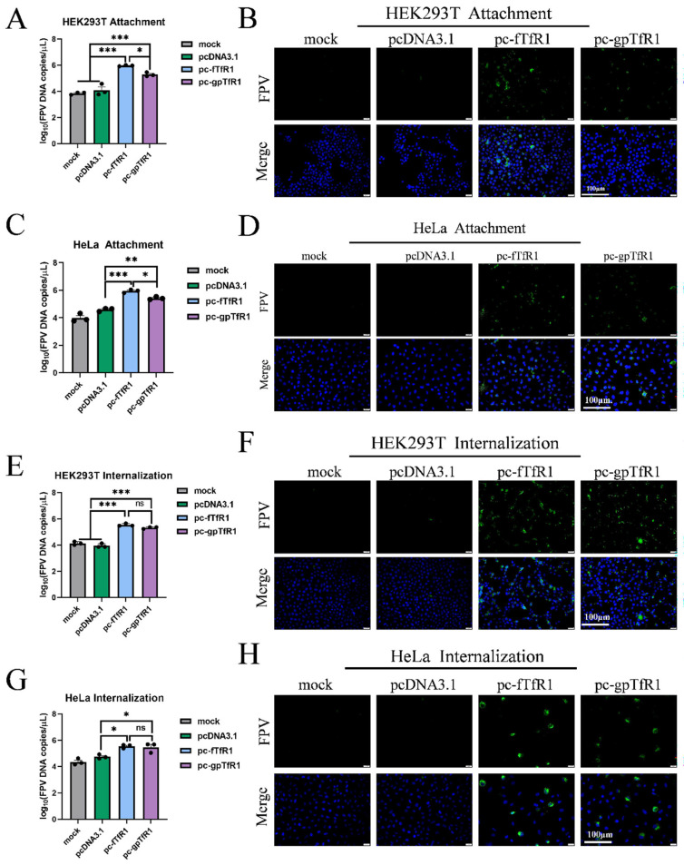
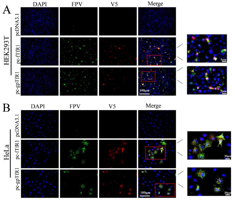
Feline parvovirus (FPV) causes feline panleukopenia, a highly contagious disease in cats, marked by severe leukopenia, biphasic fever, diarrhea, vomiting, and hemorrhagic enteritis. Recently, FPV infection in giant pandas has increased, causing diarrhea and ultimately fatal outcomes, thereby threatening their survival and reproduction. Here, we investigated the transmission of FPV in giant pandas and its interaction with cellular receptors using an FPV strain (pFPV-sc) isolated from giant panda feces. Recombinant feline transferrin receptor 1 (fTfR1) and the giant panda ortholog (gpTfR1) were expressed in non-susceptible HEK293T and HeLa cells, while viral infection levels were measured to determine the effect of gpTfR1 on pFPV-sc replication. The findings indicated that gpTfR1 overexpression in non-susceptible cells significantly enhanced pFPV-sc replication, particularly influencing the viral attachment and internalization stages. Our data further revealed early-stage colocalization between gpTfR1 expression and virus infection, suggesting that gpTfR1 facilitates early viral infection and replication. Taken together, our study provides the first evidence on the mechanism of FPV cross-species infection in giant pandas and elucidates the interaction between gpTfR1 and FPV, which establishes a theoretical basis for the development of preventive and therapeutic strategies, thereby safeguarding the health and survival of giant panda populations from FPV.

Keywords: feline parvovirus; giant panda; transferrin receptor 1; virus replication.


Conflict of interest statement

The authors declare no conflict of interest.

Figures

Figure 1
Figure 1
pFPV-sc infection in F81, HEK293T, and HeLa cells. Green fluorescence represents successful pFPV-sc infection.
Figure 2
Figure 2
Expression identification of the recombinant fTfR1 and gpTfR1 plasmids. (A,B) CCK8 assay to detect cell viability after transfection with recombinant plasmids. (C,D) qPCR to detect the mRNA level of TfR1 in cells. (E,F) Indirect immunofluorescence assay against V5 or Myc tag to detect the expression of the fTfR1 and gpTfR1 overexpression vectors. (Blue: nucleus; Green: V5 or Myc tag; *** p < 0.001).
Figure 3
Figure 3
Overexpression of gpTfR1 promotes pFPV-sc replication. (A,B) qPCR to detect FPV copies. (C,D) Indirect immunofluorescence assay to detect the number of infected cells (Blue: nucleus; Green: FPV). (E,F) ImageJ counts of the percentage of infected cells. (* p < 0.05, ** p < 0.01, *** p < 0.001).
Figure 4
Figure 4
Effect of gpTfR1 expression on the internalization stage of pFPV-sc adsorption. (A,C,E,G) FPV copy numbers detected using qPCR. (B,D,F,H) The number of infected cells detected by indirect immunofluorescence assay. (Blue: nucleus; Green: FPV; ns: not significant; * p < 0.05, ** p < 0.01, *** p < 0.001).
Figure 5
Figure 5
Immunofluorescence colocalization of gpTfR1 with pFPV-sc. (A) Colocalization of gpTfR1 and FPV in HEK293 cells. (B) Colocalization of gpTfR1 and FPV in HeLa cells. (Blue: nucleus; Green: FPV; Red: V5 tag).

Similar articles

References

    1. Jager M.C., Tomlinson J.E., Lopez-Astacio R.A., Parrish C.R., Van de Walle G.R. Small but mighty: Old and new parvoviruses of veterinary significance. Virol. J. 2021;18:210. doi: 10.1186/s12985-021-01677-y. - DOI - PMC - PubMed
    1. Wang K., Du S.S., Wang Y.Q., Wang S.Y., Luo X.Q., Zhang Y.Y., Liu C.F., Wang H.J., Pei Z.H., Hu G.X. Isolation and identification of tiger parvovirus in captive siberian tigers and phylogenetic analysis of VP2 gene. Infect. Genet. Evol. 2019;75:103957. doi: 10.1016/j.meegid.2019.103957. - DOI - PubMed
    1. Studdert M., Kelly C., Harrigan K. Isolation of panleucopaenia virus from lions. Vet. Rec. 1973;93:156–158. doi: 10.1136/vr.93.6.156. - DOI - PubMed
    1. Huang S., Li X., Xie W., Guo L., You D., Xu H., Liu D., Wang Y., Hou Z., Zeng X., et al. Molecular detection of parvovirus in captive Siberian tigers and lions in Northeastern China from 2019 to 2021. Front. Microbiol. 2022;13:898184. doi: 10.3389/fmicb.2022.898184. - DOI - PMC - PubMed
    1. Barker I.K., Povey R.C., Voigt D.R. Response of mink, skunk, red fox and raccoon to inoculation with mink virus enteritis, feline panleukopenia and canine parvovirus and prevalence of antibody to parvovirus in wild carnivores in Ontario. Can. J. Comp. Med. 1983;47:188. - PMC - PubMed

LinkOut - more resources