

Assessment of Pulmonary Vein Diameters in Cavalier King Charles Spaniels with Myxomatous Mitral Valve Disease
- PMID: 40711275
- PMCID: PMC12299408
- DOI: 10.3390/vetsci12070615
Assessment of Pulmonary Vein Diameters in Cavalier King Charles Spaniels with Myxomatous Mitral Valve Disease
Abstract
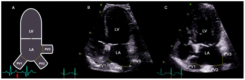
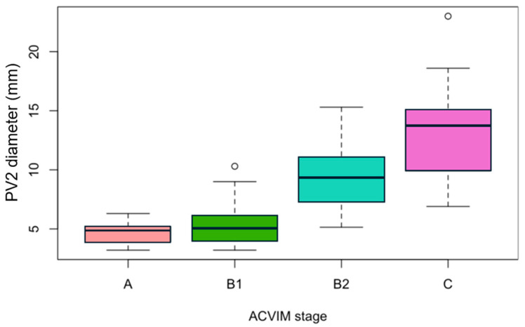
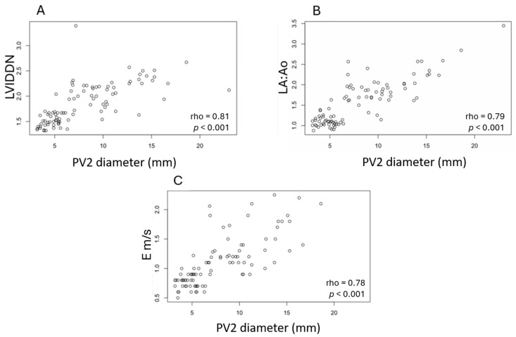
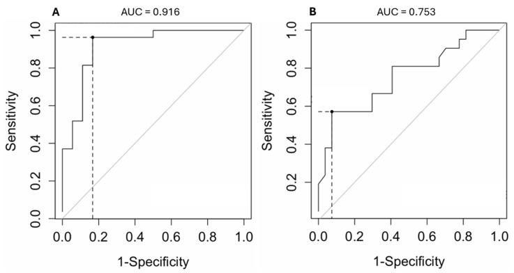
The present study aimed to compare pulmonary vein (PV) diameters between Cavalier King Charles Spaniels (CKCSs) with myxomatous mitral valve disease (MMVD) and healthy CKCSs, assess correlations between PV diameters and echocardiographic parameters, and identify the optimal PV diameter cut-off value that distinguishes stage C from stage B2. CKCSs were recruited both retrospectively and prospectively and classified according to the ACVIM guidelines (stages A, B1, B2, and C). From a left apical view, the diameters of three PVs (PV1, PV2, and PV3) were measured with high reproducibility. In healthy dogs, the PV2 diameter showed no correlation with body weight. The PV2 diameter was significantly higher in stage B2 compared to B1 and in stage C compared to B2, while no difference was found between stages A and B1. The median (IQR) PV2 diameters were 4.9 mm (3.9-5.2) in stage A, 5.1 mm (4.0-6.0) in stage B1, 9.3 mm (7.3-11.1) in stage B2, and 13.7 mm (9.9-15.1) in stage C. Positive correlations were observed between the PV2 diameter and the left ventricular internal diameter normalized for body weight, the left atrium-to-aorta ratio, mitral E wave peak velocity, tricuspid regurgitation pressure gradient, and regurgitant fraction. A PV2 diameter cut-off value of 12.8 mm discriminated stage C from stage B2 with 57% sensitivity and 93% specificity. The PV2 diameter is a reproducible echocardiographic measure that increases with MMVD severity and could assist in the early detection of congestive heart failure. However, the modest sensitivity observed reflects the overlap of PV2 measurements between stages B2 and C. Therefore, PV2 should be interpreted with caution and considered a supportive, rather than exclusive, tool in disease staging and therapeutic decision-making.
Keywords: Cavalier King Charles Spaniels; canine; congestive heart failure; myxomatous mitral valve disease; pulmonary vein.
Conflict of interest statement
The authors declare no conflicts of interest.
Figures
References
-
- Keene B.W., Atkins C.E., Bonagura J.D., Fox P.R., Häggström J., Fuentes V.L., Oyama M.A., Rush J.E., Stepien R., Uechi M. ACVIM consensus guidelines for the diagnosis and treatment of myxomatous mitral valve disease in dogs. J. Vet. Intern. Med. 2019;33:1127–1140. doi: 10.1111/jvim.15488. - DOI - PMC - PubMed
-
- Menciotti G., Borgarelli M., Aherne M., Wesselowski S., Häggström J., Ljungvall I., Lahmers S.M., Abbott J.A. Mitral valve morphology assessed by three-dimensional transthoracic echocardiography in healthy dogs and dogs with myxomatous mitral valve disease. J. Vet. Cardiol. 2017;19:113–123. doi: 10.1016/j.jvc.2017.01.002. - DOI - PubMed
-
- Boswood A., Haggstrom J., Gordon S.G., Wess G., Stepien R.L., Oyama M.A., Keene B.W., Bonagura J., MacDonald K.A., Patteson M., et al. Effect of Pimobendan in dogs with preclinical myxomatous mitral valve disease and cardiomegaly: The EPIC Study—A randomized clinical trial. J. Vet. Intern. Med. 2016;30:1765–1779. doi: 10.1111/jvim.14586. - DOI - PMC - PubMed
-
- Hansson K., Häggström J., Kvart C., Lord P. Left atrial to aortic root indices using two-dimensional and M-mode echocardiography in Cavalier King Charles Spaniels with and without left atrial enlargement. Vet. Radiol. Ultrasound. 2002;43:568–575. doi: 10.1111/j.1740-8261.2002.tb01051.x. - DOI - PubMed
LinkOut - more resources
Full Text Sources
Research Materials
