. 2025 Jun 30;12(7):628.
doi: 10.3390/vetsci12070628.

Biofilm Formation, Antibiotic Resistance, and Virulence Analysis of Human and Avian Origin Klebsiella pneumoniae from Jiangsu, China

Affiliations

Biofilm Formation, Antibiotic Resistance, and Virulence Analysis of Human and Avian Origin Klebsiella pneumoniae from Jiangsu, China

Yulu Xue et al. Vet Sci. .

Abstract

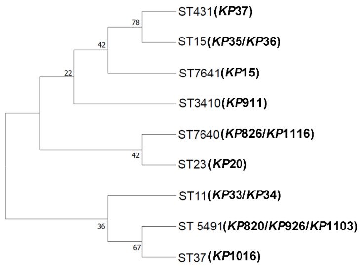
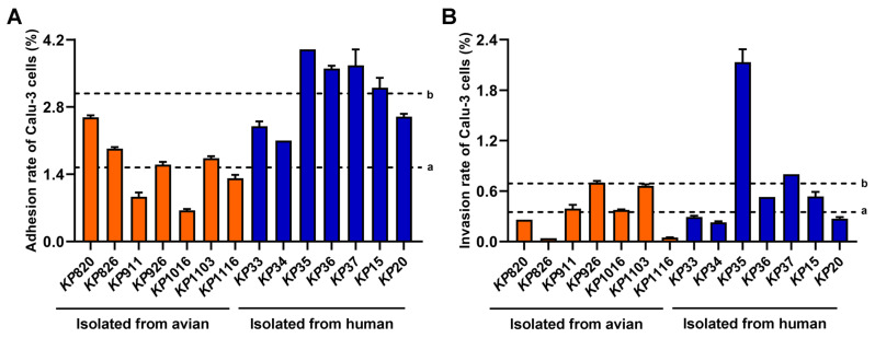
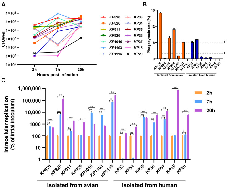
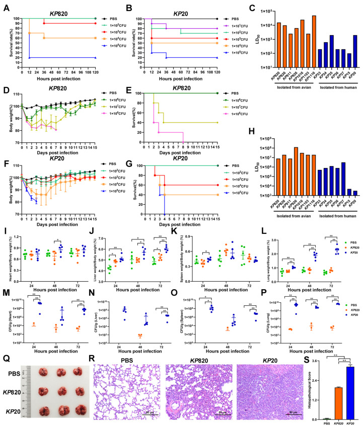
Klebsiella pneumoniae, a zoonotic pathogen of global concern, poses significant threats to both veterinary and public health. Here, a comparative study characterized 14 clinical isolates (7 avian-derived, 7 human-derived) from Jiangsu, China, through integrated genomic and phenotypic analyses. Firstly, multilocus sequence typing (MLST) revealed distinct epidemiological patterns: the same ST type in avian isolates was circulating between different species and different regions, whereas it was not found in human isolates. In addition, hypervirulent Klebsiella pneumoniae (hvKP) phenotypes confirmed by string test were exclusive to two human isolates (KP15, KP20). Secondly, biofilm detection demonstrated 78.6% (11/14) of isolates possessed biofilm-forming capacity, with cellulose but not curli as the predominant matrix component. Human-derived KP15 and KP20 had the strongest biofilm formation ability in all isolates. Antimicrobial susceptibility profiling identified serious multidrug resistance in both avian and human isolates. Virulence gene analysis revealed striking disparities, with human isolates harboring 10-20 virulence factors (median 15) versus 6-7 (median 6.5) in avian counterparts. Finally, functional pathogenesis assessments demonstrated human-derived strains exhibited stronger epithelial cell adhesion (2-fold higher) and invasion (1.97-fold higher) in Calu-3 cell models and paradoxically showed reduced macrophage phagocytosis (2.85-fold lower at 2 h) for immune escape. In vivo models confirmed dose-dependent mortality, with human isolates demonstrating higher lethality in both Galleria mellonella and mice. Virulence gene burden positively correlated with mortality outcomes. These findings delineate critical host adaptation differences in Klebsiella pneumoniae populations and provide empirical evidence for pathogen transmission dynamics at the human-animal interface.

Keywords: Klebsiella pneumoniae; antibiotic resistance; avian-origin; biofilm; human-origin; virulence.


Conflict of interest statement

The authors have no financial conflicts of interest.

Figures

Figure 1
Figure 1
Neighbor-joining phylogeny was constructed using seven MLST genes for 14 K. pneumoniae isolates with 1000 bootstrap replicates. The high bootstrap values implied a high level of confidence in the tree.
Figure 2
Figure 2
Results of viscosity assay. (A,B) KP15 and KP20 could form a string on the LB agar plate. Generally, a string 5 mm or longer was defined as positive. *, the distance from the bottom of the toothpick to the black spot is 5 mm. (C,D) KP826 and KP911 could not form a string on the LB agar plate. (E) Mucoviscosity assay of K. pneumoniae isolates. (F) The bacteria were evenly mixed on the slide with Congo red and serum mixture solution and then were pushed into a thin layer. The bacteria were decolonized with 5% dilute hydrochloric acid for 1 min, washed with water, and then stained with 0.1% crystal violet for 1 min, washed with water, dried naturally, and observed under the oil microscope. Bars: 10 μm. Capsules are indicated by short red arrows.
Figure 3
Figure 3
Results of biofilm formation ability of K. pneumoniae isolates. (A) The biofilm formation was detected by the crystal violet in the test tube. (B) The crystal violet was dissolved using absolute ethanol, and the absorbance was measured at a wavelength of 550 nm. (C) Biofilm structure of KP826 and KP15 was observed by confocal laser scanning microscopy (CLSM). (D) Colony morphology of 14 K. pneumoniae isolates growing on Congo red plates. (E) Colony morphology of 14 K. pneumoniae isolates growing on Calcofluor plates. Photographs represent one of three experiments, which gave similar results.
Figure 4
Figure 4
Result of epithelial cell adhesion and invasion assay. (A) Adhesion ability of K. pneumoniae to Calu-3 cells. (B) Invasion ability of K. pneumoniae to Calu-3 cells. a: The average adhesion rate or invasion rate of strains isolated from avian to Calu-3 cells. b: The average adhesion rate or invasion rate of strains isolated from humans to Calu-3 cells.
Figure 5
Figure 5
Results of K. pneumoniae infection in macrophage RAW264.7. (A) The amount of K. pneumoniae strains inside macrophage RAW264.7 at 2, 7, and 20 h after infection, respectively. (B) Phagocytosis rate of macrophages RAW264.7 against K. pneumoniae. a: The average phagocytosis rate of macrophages to strains isolated from avian. b: The average phagocytosis rate of macrophages to strains isolated from humans. (C) The intracellular replication rates of K. pneumoniae strains inside macrophage RAW264.7. The intracellular replication at 7 and 20 h was calculated by comparing to the CFUs at 2 h *, p < 0.05; **, p < 0.01.
Figure 6
Figure 6
Evaluation of pathogenicity of K. pneumoniae isolates. (A) Survival rate of Galleria mellonella (n = 10) infected with KP820. (B) Survival rate of Galleria mellonella (n = 10) infected with KP20. (C) K. pneumoniae strains infected Galleria mellonella, then calculated median lethal dose. (D,E) The body weight changes and survival rate of mice (n = 5) intratracheally injected with KP820. (F,G) The body weight changes and survival rate of mice (n = 5) intratracheally injected with KP20. (H) K. pneumoniae strains infected mice, then calculated the median lethal dose. (IL) The wet weight of the tissue (heart, liver, spleen, and lung) in mice (n = 5) intratracheally injected with PBS, KP820, and KP20 at 24, 48, and 72 h post infection. (MP) The bacterial load of the tissue (heart, liver, spleen, and lung) in mice (n = 5) intratracheally injected with PBS, KP820, and KP20 at 24, 48, and 72 h post infection. The pathology images in lungs (n = 3 per group) (Q), histopathological images in H&E-stained lung tissues (n = 3) (R), and histopathologic scores in the lungs (S) of mice intratracheally injected with PBS, KP820, and KP20 at 72 h post infection. *, p < 0.05; **, p < 0.01.

References

    1. Liang J., Lin S., He L. Formation ability and drug resistance mechanism of Klebsiella pneumoniae biofilm and capsule for multidrug-resistant. Cell. Mol. Biol. 2023;69:88–93. doi: 10.14715/cmb/2023.69.10.12. - DOI - PubMed
    1. Riwu K.H.P., Effendi M.H., Rantam F.A., Khairullah A.R., Widodo A. A review: Virulence factors of Klebsiella pneumonia as emerging infection on the food chain. Vet. World. 2022;15:2172–2179. doi: 10.14202/vetworld.2022.2172-2179. - DOI - PMC - PubMed
    1. Bina X.R., Weng Y., Budnick J., Van Allen M.E., Bina J.E. Klebsiella pneumoniae TolC contributes to antimicrobial resistance, exopolysaccharide production, and virulence. Infect. Immun. 2023;91:e0030323. doi: 10.1128/iai.00303-23. - DOI - PMC - PubMed
    1. Shao Y., Wang Y., Yuan Y., Xie Y. A systematic review on antibiotics misuse in livestock and aquaculture and regulation implications in China. Sci. Total Environ. 2021;798:149205. doi: 10.1016/j.scitotenv.2021.149205. - DOI - PubMed
    1. Mobasseri G., Teh C.S.J., Ooi P.T., Thong K.L. The emergence of colistin-resistant Klebsiella pneumoniae strains from swine in Malaysia. J. Glob. Antimicrob. Resist. 2019;17:227–232. doi: 10.1016/j.jgar.2018.12.015. - DOI - PubMed

LinkOut - more resources