. 2025 Jul 31;39(14):e70879.
doi: 10.1096/fj.202501431RR.

Omicron Impacts Olfaction in Hamsters

Affiliations

Omicron Impacts Olfaction in Hamsters

Virginie Guérin et al. FASEB J. .

Abstract

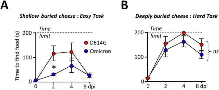
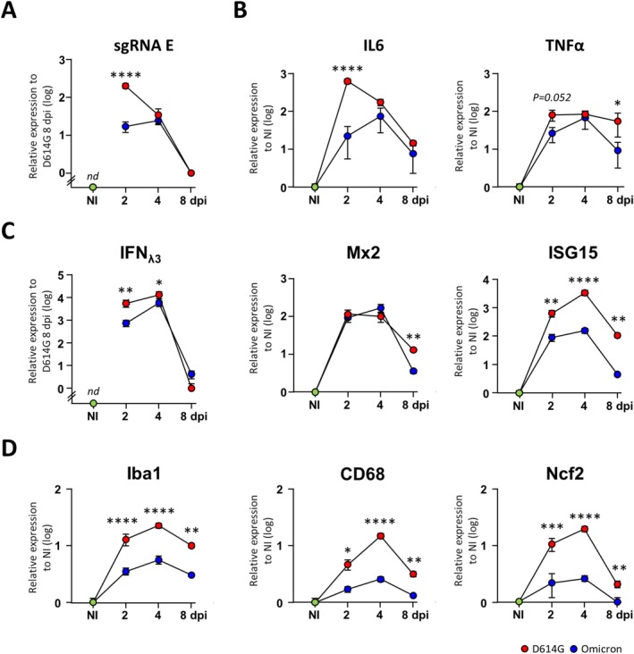
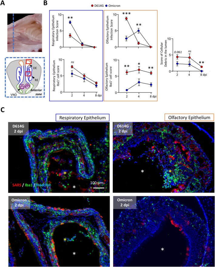
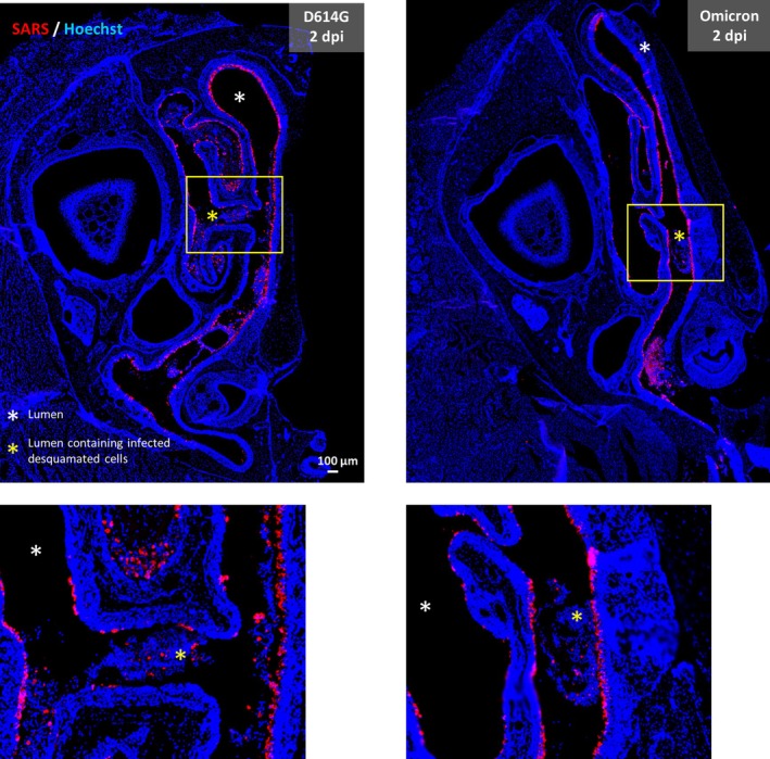
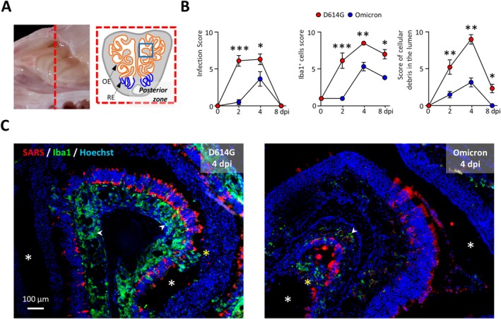
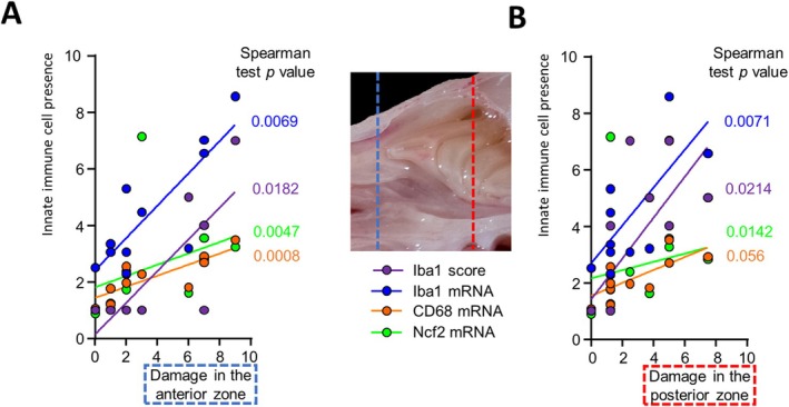
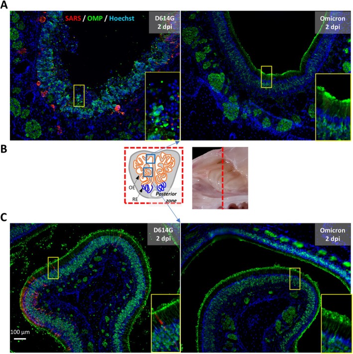
Olfactory disorders have been a hallmark of COVID-19. Since the emergence of Omicron, the prevalence of anosmia has dropped, reducing interest for Omicron pathophysiology in the nasal cavity. Using the hamster model, we show that despite a lower impact than early variants of SARS-CoV-2 (D614G), Omicron BA.1 infection leads to hyposmia as early as 2 days postinfection (dpi). While the olfactory epithelium was mostly spared from Omicron infection at 2 dpi, the respiratory epithelium was strongly infected with increased presence of inflammation markers, epithelial damage, and cellular debris in the lumen of the nasal cavity. The hyposmia caused by the early SARS-CoV-2 variant and Omicron BA.1 infection was similar at 4 and 8 dpi. At 4 dpi, Omicron successfully infected the olfactory epithelium although to a lesser extent than D614G. The BA.1 infection led to innate immune cell invasion and desquamation of the olfactory epithelium similarly as the D614G variant, with persistent inflammatory markers in the olfactory turbinates at 8 dpi despite clearance of the virus. Overall, our results indicate that Omicron successfully infects the nasal epithelium including the olfactory epithelium, but with a delay and to a lesser extent than previous SARS-CoV-2 variants. These results are consistent with Omicron pathophysiology in humans showing a decreased olfactory threshold sensitivity that may be caused, as at the early stage in hamsters, by cellular debris filling the lumen of the olfactory cleft following epithelial damage.

Keywords: olfaction; olfactory behavior; respiratory viruses.


Conflict of interest statement

The authors declare no conflicts of interest.

Figures

FIGURE 1
FIGURE 1
Olfactory abilities of SARS‐CoV‐2‐infected hamsters are impacted by Omicron variant, but to a lesser extent than D614G VoC. The amount of time taken to find a shallow (A) or deeply (B) buried piece of cheese (mean ± SEM, n = 6, two‐way ANOVA followed by Bonferroni's multiple comparisons test; ns, nonsignificant; *p < 0.01). The animal has 200 s to find the food before the test ends. s, second.
FIGURE 2
FIGURE 2
Comparative kinetics of gene expression related to virus and host response following D614G and Omicron infection in hamster olfactory turbinates. Evolution of gene expression related to (A) viral subgenomic RNA E, (B) inflammatory cytokines, (C) interferon pathway, and (D) innate immune cells (Iba1 and CD68 for resident and circulating macrophages, respectively; Ncf2 for neutrophils). NI, non‐infected controls. Relative expression mean ± SEM, n = 6, Mann–Whitney test; *p < 0.05; **p < 0.01; ***p < 0.001; ****p < 0.0001.
FIGURE 3
FIGURE 3
Omicron is more present in the respiratory epithelium than D614G and infects the olfactory epithelium with a delay in the anterior zone of the nasal cavity. (A) Localization of the anterior zone in the nasal cavity. (B) Score of infection, of resident macrophage presence, and of cellular debris filling the lumen of nasal cavity in the respiratory and olfactory epithelium. Mean ± SEM, n = 6, Mann–Whitney test; *p < 0.05; **p < 0.01; ***p < 0.001. (C) Representative images of respiratory (RE) and olfactory epithelium (OE) in hamsters infected with either D614G or Omicron BA.1 VoCs at 2 dpi (SARS‐CoV‐2 infected cells in red; infiltrated resident macrophages in green; lumen of the nasal cavity—white asterisk—filled with cellular debris and infiltrated immune cells—yellow asterisk). Localization of images is indicated by the red squares in panel A. VNO, vomeronasal organ.
FIGURE 4
FIGURE 4
Presence of cellular debris in the lumen of the nasal cavity at 2 dpi following D614G and Omicron infection. Representative image of the anterior zone of the hamster nasal cavity following infection with D614G (left) or BA.1 Omicron (right) VoCs (SARS‐CoV‐2 infected cells in red). Note the presence of infected cellular debris in the lumen of the nasal cavity.
FIGURE 5
FIGURE 5
Omicron infection does not spare the olfactory epithelium in the posterior zone of the nasal cavity. (A) Localization of the posterior zone of the nasal cavity. (B) Score of infection; Iba1+ cells (resident macrophages) in the olfactory epithelium and cellular debris filling the lumen of the nasal cavity. Mean ± SEM, n = 6, Mann–Whitney test; *p < 0.05; **p < 0.01; ***p < 0.001. (C) Representative images of the olfactory epithelium in hamsters infected by either D614G or Omicron BA.1 VoCs at 2 dpi (SARS‐CoV‐2 infected cells in red; infiltrated resident macrophages in green—indicated by a white arrow head in the lamina propria; lumen of the nasal cavity—white asterisk—filled with cellular debris and infiltrated immune cells—yellow asterisk). The location of images is indicated by the blue square in panel A.
FIGURE 6
FIGURE 6
Innate immune cell presence is related to OE damage following BA.1 infection. Correlation between damage in the olfactory epithelium (OE) and the presence of innate immune cells. Resident macrophages were measured by Iba1+ immunohistochemistry or Iba1 gene expression level; circulating macrophages and neutrophils by CD68 and Ncf2 mRNA expression levels, respectively (n = 12 from 2 and 4 dpi BA.1 infected hamsters) in the (A) anterior or (B) posterior zone of the nasal cavity.
FIGURE 7
FIGURE 7
Differential impact of D614G and BA.1 Omicron infection on olfactory sensory neurons integrity in the posterior zone of the nasal cavity at 2 dpi. (A) Dorsal OE infected with D614G (left side) and Omicron (right side). (B) Location of images is indicated by the blue squares. (C) Intermediate OE infected with D614G (left side) but not with Omicron (right side).

Similar articles

References

    1. Planas D., Saunders N., Maes P., et al., “Considerable Escape of SARS‐CoV‐2 Omicron to Antibody Neutralization,” Nature 602 (2022): 671–675. - PubMed
    1. Chen M., Pekosz A., Villano J. S., et al., “Evolution of Nasal and Olfactory Infection Characteristics of SARS‐CoV‐2 Variants,” Journal of Clinical Investigation 134, no. 8 (2024): e174439. - PMC - PubMed
    1. Shuai H., Chan J. F.‐W., Hu B., et al., “Attenuated Replication and Pathogenicity of SARS‐CoV‐2 B.1.1.529 Omicron,” Nature 603 (2022): 693–699. - PubMed
    1. Halfmann P. J., Iida S., Iwatsuki‐Horimoto K., et al., “SARS‐CoV‐2 Omicron Virus Causes Attenuated Disease in Mice and Hamsters,” Nature 603 (2022): 687–692. - PMC - PubMed
    1. von Bartheld C. S. and Wang L., “Prevalence of Olfactory Dysfunction With the Omicron Variant of SARS‐CoV‐2: A Systematic Review and Meta‐Analysis,” Cells 12 (2023): 430. - PMC - PubMed

Supplementary concepts

LinkOut - more resources