. 2025 Jul 25;82(1):287.
doi: 10.1007/s00018-025-05781-y.

Fibroblasts activated by miRs-185-5p, miR-652-5p, and miR-1246 shape the tumor microenvironment in triple-negative breast cancer via PATZ1 downregulation

Affiliations

Fibroblasts activated by miRs-185-5p, miR-652-5p, and miR-1246 shape the tumor microenvironment in triple-negative breast cancer via PATZ1 downregulation

Giada De Luca et al. Cell Mol Life Sci. .

Abstract

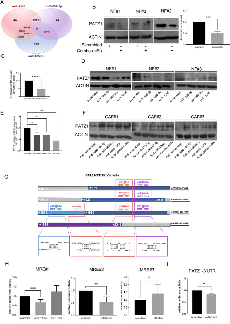
The intricate interplay between epithelial and fibroblast cells within the tumor microenvironment plays a crucial role in driving triple-negative breast cancer progression. This crosstalk involves the exchange of various signaling molecules, including growth factors, cytokines, extracellular matrix components, and extracellular vesicles. Recently, we demonstrated that triple-negative breast cancer extracellular vesicles carry and release a specific combination of miRs, including miR-185-5p, miR-652-5p, and miR-1246 (from here on, referred as combo-miRs), into normal fibroblasts, effectively reprogramming them into cancer-associated fibroblasts. Here, we show that the conditioned medium from the fibroblasts activated by combo-miRs exerts a pro-tumorigenic effect on epithelial cells, enhancing the viability and migratory potential while driving increased invasiveness in patient-derived breast cancer organoids. A proteomic analysis of conditioned medium from combo-miRs activated fibroblasts revealed 76 significantly upregulated secreted proteins compared to control. Bioinformatic analysis identified the transcriptional factor PATZ1 as a potential regulator of the 12 most highly upregulated proteins. Consistently, in-silico predictions and in vitro experiments confirmed that PATZ1 is a direct target of miR-185-5p and miR-652-5p. The downregulation of PATZ1 by these miRNAs led to increased levels of the secreted proteins in the conditioned medium from combo-miRs activated fibroblasts. Furthermore, the conditioned medium from PATZ1-knockout mesenchymal embryonic fibroblasts and normal fibroblasts with silenced PATZ1 similarly enhanced the migratory potential of MCF10A cells, further supporting the critical role of PATZ1 in regulating tumor-promoting mechanisms. These findings provide valuable insights into the dynamics of the TME in TNBC, highlighting combo-miRs and PATZ1 as promising targets for future therapeutic interventions.

Keywords: CAFs; Extracellular vesicles; Fibroblasts; PATZ1; miRNAs.


Conflict of interest statement

Declarations. Competing interests: Alessandra Affinito and Sara Verde are employees of AKA Biotech SRL. However, this employment did not influence the design, execution, or interpretation of the research presented in this manuscript. The other authors declare no other competing interests.

Figures

Fig. 1
Fig. 1
Conditioned medium from combo-miRs-activated fibroblasts increases epithelial breast aggressiveness. A MTS assay performed on luminal A-MCF7 and non-tumorigenic-MCF10A breast cells pre-treated with CM from NFs (pt. #1, #2) transfected for 72 h with combo miRs. The absorbance at 492 nm was measured after 48 h (t48), normalized to t0, and folded over the control. Data are shown as two biological replicates B Migration assay of MCF10A cells evaluated using CM_combo miRs/CM_Scra (pt. #1, #2, #3) as a chemoattractant. Representative images were captured with phase-contrast microscopy after staining with crystal violet. The histogram reports the absorbance at 595 nm of the SDS-eluted crystal violet. Data are shown as two biological replicates. One-way Anova test C Representative optical microscope images (20x magnification) of luminal A breast cancer organoids (pt. # 535) embedded in type 1 collagen matrix and incubated with CM_pt#1 Combo miRs and CM_pt#1 Scra (control) captured at 0, 24, 48, 72, and 96 h. CM was added at 0 h and 48 h (as indicated by the red arrows). White lines delimit invasive protrusions surrounding the organoids. Red arrows indicate the supplement of the CM. Pixel bars represent the scale. The graph displays the quantification of organoid invasion obtained by measuring the area of organoids at 72 and 96 h, normalized to t0, and compared to the control. Image J software was used to measure the area of protrusions. The standard deviation was determined using technical replicates. The adjusted p-value was obtained with 2-way ANOVA. ** P values < 0.01, ***p < 0.001, **** P values < 0.0001
Fig. 2
Fig. 2
Differential proteome profile in conditioned medium from combo-miRs activated fibroblasts compared to control. A Representative volcano plot of upregulated proteins CM_combo-miRs vs. control. All the proteins shown in the graph are significantly upregulated in CM from combo-miRs activated fibroblasts (CM_combo-miRs) compared to the control (CM_scra), as indicated by blue and black points. B Heatmap of proteomic analysis demonstrating proteins differently expressed in CM_combo-miRs compared to CM_scra. Downregulated proteins are represented in red, and upregulated proteins are presented in green. C Most upregulated proteins (FC>2) were subjected to functional analysis using GO (C-D), wikiPathways E and Reactome F databases. Results showed that the genes were enriched in processes related to extracellular matrix organization, cellular migration, and ECM protein binding. G ORA signature analysis on all Differently Expressed proteins (log2 fold change < −0.5 & > 0.5), From the results datatable we extracted all the terms related to ‘fibrolasts’. The analysis showed that combo-miR-activated fibroblasts belong to the Myofibroblasts phenotypic class. H ELISA assay on CM_combo-miRs to validate the proteomic analysis. Graphs show mean expression levels of Vimentin, Sirp alpha, TGF β1, SRPX, in pt. #2, #3, #4, #5 (fold on CM_scra). Data are calculated as three technical replicates of four biological replicates. Unpaired t-test. *P values <0.05, ** P values < 0.01
Fig. 3
Fig. 3
PATZ1 regulates EMT-secreted protein in CMs. A Real-time PCR of Vimentin, Sirp alpha, TGF β1, SRPX and RAB1B, in BT549 cell lines upon HA-PATZ1 overexpression. Data are calculated as two technical replicates of experimental triplicates B Migration assay of MCF7 cells with CM_MEF +/- or +/+ and CM_wild type MEF (NT) as a chemoattractant. Representative images were captured with phase-contrast microscopy after staining with crystal violet. The histogram reports the absorbance at 595 nm of the SDS-eluted crystal violet. ANOVA test calculated the p-value on triplicates of two independent experiments. C Scratch assay of MCF7 cells evaluated using CM_MEF +/- or +/+ and CM_wildtype MEF (NT) as a chemoattractant. Representative images were captured with phase-contrast microscopy at time 0 and 48 h after treatment. The histogram on the right reports measured area upon wound healing normalized on the t0 areas. Standard deviations were calculated on replicates from two independent experiments performed on MCF7. Anova test calculated the p-value D Scratch assay of MCF7 cells with CM_PATZ1 siRNA and CM_NT as a chemoattractant on three different patients (#1, #2, #3). Representative images were captured with phase-contrast microscopy at time 0 and 48 h after treatment. The histogram on the right reports measured area upon wound healing normalized on the t0 areas. Standard deviations were calculated on biological replicates of three patient’s derived CMs performed on MCF7. ANOVA test calculated the p-value (** p < 0.01, ** p < 0.0001). E Migration assay of MCF7 cells with CM_PATZ1 siRNA and CM_NT as a chemoattractant. Representative images were captured with phase-contrast microscopy after staining with crystal violet. The histogram reports the absorbance at 595 nm of the SDS-eluted crystal violet. ANOVA test calculated the p-value on biological replicates of five patient’s derived CMs performed on MCF7 (* p < 0.05, ** p < 0.01). F MTT assay of MCF7 cells treated with CM_MEF +/- or +/+ and CM_wildtype MEF (NT) for 6 days. Data are reported as fold on NT of two independent experiments. * p < 0.05. *P values < 0.05, ** P values < 0.01, ***p < 0.001, **** P values < 0.0001
Fig. 4
Fig. 4
PATZ1 is a target of miR-185-5p, miR-652-5p and miR-1246. A Venn diagram showing the intersection of target genes of miR-185-5p, miR-652-5p, and miR-1246 predicted by the miRDB database. PATZ1 was identified as a common target of miR-185-5p and miR-652-5p. B Western blot analysis of PATZ1 protein upon combo-miRs overexpression in NFs from three different patients. B-Actin was used as a loading control. C Real-time PCR of PATZ1 mRNA upon combo-miRs upregulation. Student t-test was calculated on four independent experiments. D Western blot analysis of PATZ1 protein upon miR-185-5p, miR-652-5p and miR-1246 overexpression in NFs from three patients. B-Actin was used as a loading control. E Real-time PCR of PATZ1 mRNA upon miR-185-5p, miR-652-5p and miR-1246 upregulation on two NFs as assessed by one way ANOVA on biological replicates of two patients. F Western blot analysis of PATZ1 protein upon Anti-miR-185-5p, Anti-miR-652-5p, Anti-miR-1246, and anti-combo-miR overexpression in NFs from three different patients. B-Actin was used as a loading control. G Schematic representation of PATZ1 3’UTR and potential miRNA target sites. H Luciferase activity of cloned MRE in pmiRGlo. Data were normalized on scrambled sequences. Student t-test was calculated on three technical replicates of two independent experiments. I Luciferase activity of hs-PATZ1-3’UTR v202-203 cloned in pgl3 vector. Data were normalized on scrambled sequences. Student t-test was calculated on three technical replicates of two independent experiments. ** p < 0.01

References

    1. Nolan E, Lindeman GJ, Visvader JE (2023) Deciphering breast cancer: from biology to the clinic. Cell 186:1708–1728. 10.1016/j.cell.2023.01.040 - PubMed
    1. Rivenbark AG, O’Connor SM, Coleman WB (2013) Molecular and cellular heterogeneity in breast cancer: challenges for personalized medicine. Am J Pathol 183:1113–1124. 10.1016/j.ajpath.2013.08.002 - PMC - PubMed
    1. Farkas AH, Nattinger AB (2023) Breast Cancer screening and prevention. Ann Intern Med 176:ITC161–ITC. 10.7326/AITC202311210 - PubMed
    1. Patel G, Prince A, Harries M (2024) Advanced Triple-Negative breast Cancer. Semin Oncol Nurs 40:151548. 10.1016/j.soncn.2023.151548 - PubMed
    1. Capuozzo M, Celotto V, Santorsola M, Fabozzi A, Landi L, Ferrara F, Borzacchiello A, Granata V, Sabbatino F, Savarese G, Cascella M, Perri F, Ottaiano A (2023) Emerging treatment approaches for triple-negative breast cancer. Med Oncol 41:5. 10.1007/s12032-023-02257-6 - PubMed

MeSH terms

LinkOut - more resources