. 2025 Jul 25;11(30):eadw3307.
doi: 10.1126/sciadv.adw3307. Epub 2025 Jul 25.

Deep homology of a brachyury cis-regulatory syntax and the evolutionary origin of the notochord

Affiliations

Deep homology of a brachyury cis-regulatory syntax and the evolutionary origin of the notochord

Tzu-Pei Fan et al. Sci Adv. .

Abstract

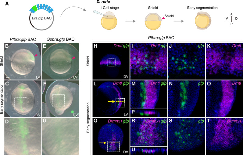
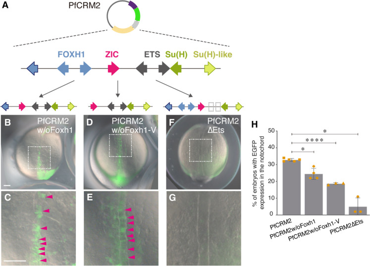
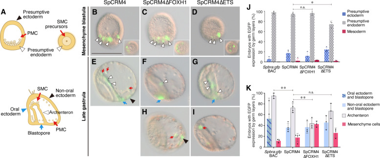
Expression of brachyury in the notochord is regarded as a chordate novelty and links to the origin of the notochord, yet the evolution of this regulatory control remains unclear. Here, we uncovered a regulatory syntax (named SFZE) consisting of binding sites for four transcription factors in notochord enhancers of chordate brachyury genes. SFZE was also identified in potential brachyury enhancers in various non-chordate animals and even in Capsaspora, a unicellular relative to animals. These non-chordate SFZE-containing enhancers exhibited activity in the zebrafish notochord. Furthermore, the SFZE syntax in a non-chordate confers endoderm activity. Our results indicate the ancient association of SFZE with brachyury, likely predating the origin of animals. The emergence of notochordal brachyury expression could be attributed to co-option of upstream signals acting on the conserved SFZE syntax, which facilitates the origin of the notochord from rudimentary endodermal cells.


Figures

Fig. 1.
Fig. 1.. Pfbra:gfp BAC activates GFP expression in the organizer and the hypochord of zebrafish embryos.
(A) Circular Pfbra/Spbra:gfp BACs were each introduced into zebrafish zygotes, and GFP signals were observed at the shield and early segmentation stages (A, anterior; P, posterior; V, ventral; D, dorsal). (B to G) GFP signals in zebrafish embryos injected with Pfbra:gfp BAC or Spbra:gfp BAC. GFP signal in the dorsal margin (shield) at the shield stage is indicated by the red arrowhead. The white dashed boxes in (C) and (F) are magnified and shown in (D) and (G), respectively. (H to U) Double fluorescent in situ hybridization of gfp (green) and Drntl [magenta in (H) to (P)] or Drmnx1 (magenta in Q to U) in Pfbra:gfp BAC-injected zebrafish embryos at indicated stages. Nuclei were counterstained with Hoechst 33342 (blue). The areas in the white dashed boxes in (H), (L), and (Q) are enlarged and shown in (I), (M), and (R); single-channel images are shown in (J) and (K), (N) and (O), and (S) and (T), respectively. [(P) and (U)] The XZ sections along the yellow dashed lines are indicated with yellow arrows in (L) and (Q). The orientation of embryos is indicated in each panel: LV, lateral view; DV, dorsal view. All scale bars represent 100 μm. Panels that are in the same scale: (B), (C), (E), and (F); (D) and (G); (H), (L), and (Q); (I) to (K), (M) to (P), and (R) to (U). Created in BioRender. Fan, T. (2025) https://BioRender.com/n8z3595.
Fig. 2.
Fig. 2.. Brachyury CRMs with SFZE/SFZE-like syntax show activity in the zebrafish notochord.
(A) Phylogenetic relationships of the analyzed species. (B to Q) Zebrafish embryos injected with SFZE/SFZE-like containing CRMs from zebrafish (Drntl-1kb), hemichordate (PfCRM2), sea urchin (SpCRM4), fruit fly (DmCRM1), sea anemone (NvCRM1, NvCRM2, and NvCRM3), and filasterea (CoCRM4). The right panels show magnified views of the notochord region (white dashed boxes in the corresponding left panels) with magenta arrowheads indicating enhanced GFP (EGFP) signals in the notochord cells. The left panels are in the same scale, as are the right panels. Scale bars in (B) and (C), 100 μm. (R to W) The putative CRMs based on published ATAC-seq (Drntl, Pfbra, and Spbra loci) and chromatin immunoprecipitation sequencing (ChIP-seq) datasets (Dmbyn, H3K4me1, and H3K27me3; Nvbra, H3K4me1; Cobra, H3K4me1, and H3K4me3). Potential CRMs are indicated with numbers, and CRMs carrying SFZE/SFZE-like motifs are shaded in pink. The TF binding sites are denoted schematically with colored arrows placed on top of each diagram to show the orientation and order. Colors of TF sites are shown in (R) and (S). Genome versions, scaffold/chromosome numbers, and positions of the genomic loci displayed in (R) to (W) are indicated below the respective panel. The samples used for generating of the ATAC-seq and ChIP-seq datasets are listed in table S7. (X) Percentages of embryos with EGFP signals in the notochord out of EGFP-positive embryos. Each colored square represents the result of one experiment. Gray columns represent the average of at least three biological replicates, with error bars serving as SDs.
Fig. 3.
Fig. 3.. Foxh1 and Ets sites of the hemichordate SFZE syntax are functionally important for notochord activity.
(A) Diagram of the reporter constructs of PfCRM2 with deletions of the Foxh1 sites or mutations of the Ets sites. The additional Foxh1 site in the vector is indicated by the leftmost blue arrow with black outline. (B to G) Representative images show zebrafish embryos injected with the truncated or mutated PfCRM2 reporter constructs. Magnifications of the notochord regions highlighted by white boxes in (B), (D), and (F) are shown in (C), (E), and (G), respectively. EGFP signals in the notochord cells are indicated with magenta arrowheads. Embryos are oriented in the dorsal view with anterior to the top. Top panels [(B), (D), and (F)] and bottom panels [(C), (E), and (G)] are in the same scale as indicated in (B) and (C), respectively. w/o, without; delta symbol (△), mutation. All scale bars represent 100 μm. (H) Percentages of zebrafish embryos exhibiting EGFP signals in the notochord among EGFP-positive embryos. Each yellow data point represents the result of a single experiment. The gray columns are average results from at least three biological replicates, with error bars showing SDs. *P < 0.05; ****P < 0.0001.
Fig. 4.
Fig. 4.. Foxh1 and Ets sites of the sea urchin SFZE syntax confer endodermal activity at different developmental stages.
(A) Illustrations of sea urchin embryos at the mesenchyme blastula (top left, lateral view; top right, vegetal view) and late gastrula (bottom) stages. Colored arrows and arrowheads mark different embryonic territories. (B to I) Representative EGFP-positive embryos showing reporter activities. Colored arrows and arrowheads indicate EGFP-positive cells within the territories shown in (A). Embryos are oriented as in (A). For late gastrulae, the ventral side is to the left. Insets in (B) to (D) are the vegetal view to show EGFP signals in the presumptive endoderm. delta symbol (△), mutation. Panels are all in the same scale [scale bar in (B) 100 μm]. (J and K) Percentages of mesenchyme blastula (J) and late gastrula (K) embryos showing EGFP signals in the indicated territories. PMC, primary mesenchyme cells; SMC, secondary mesenchyme cells. *P < 0.05; **P < 0.01; n.s., not significant.
Fig. 5.
Fig. 5.. Proposed scenario of SFZE evolution and the origin of the notochord.
The SFZE syntax (red rectangles) associated with brachyury orthologs could have already been present before the emergence of animals. The regulatory network that activates brachyury in the axial mesoderm by concurrent Nodal and FGF signals through the SFZE syntax may have been established in the chordate lineage, at least before the divergence of tunicates and vertebrates. The Ets and Fox family factors (activated by yet to be determined signaling pathways, indicated by question marks) likely regulate brachyury expression through SFZE in at least ambulacrarians. Some components of the regulatory inputs could have been modified due to the loss of nodal and/or foxh genes in Drosophila and ambulacrarians (cross symbols). After two rounds of whole-genome duplications (2R), the SFZE syntax remained associated with both brachyury paralogs. Other notochord enhancers (T3/C/I) were subsequently evolved to control one of the paralogs (i.e., tbxb). In tetrapods, only tbxb was retained, and the notochord activity of the mammalian SFZE may have been substituted by T3/C/I enhancers. Created in BioRender. Fan, T. (2025) https://BioRender.com/14o3zg1.

References

    1. Stemple D. L., Structure and function of the notochord: An essential organ for chordate development. Development 132, 2503–2512 (2005). - PubMed
    1. Satoh N., Tagawa K., Lowe C. J., Yu J.-K., Kawashima T., Takahashi H., Ogasawara M., Kirschner M., Hisata K., Su Y.-H., Gerhart J., On a possible evolutionary link of the stomochord of hemichordates to pharyngeal organs of chordates: Stomochord and Notochord Relationship. Genesis 52, 925–934 (2014). - PMC - PubMed
    1. Lauri A., Brunet T., Handberg-Thorsager M., Fischer A. H. L., Simakov O., Steinmetz P. R. H., Tomer R., Keller P. J., Arendt D., Development of the annelid axochord: Insights into notochord evolution. Science 345, 1365–1368 (2014). - PubMed
    1. Hejnol A., Lowe C. J., Animal evolution: Stiff or squishy notochord origins? Curr. Biol. 24, R1131–R1133 (2014). - PubMed
    1. Bruce A. E. E., Winklbauer R., Brachyury in the gastrula of basal vertebrates. Mech. Dev. 163, 103625 (2020). - PubMed

LinkOut - more resources