. 2025 Jul 25;11(30):eadu9945.
doi: 10.1126/sciadv.adu9945. Epub 2025 Jul 25.

Structural analysis of HER2-trastuzumab complex reveals receptor conformational adaptation

Affiliations

Structural analysis of HER2-trastuzumab complex reveals receptor conformational adaptation

Santiago Vacca et al. Sci Adv. .

Abstract

Human epidermal growth factor receptor-2 (HER2) is a receptor tyrosine kinase, associated with a variety of malignant tumors, usually through overexpression, resulting in aberrant signaling. Trastuzumab (TZB), one of the monoclonal antibodies (mAbs) used in combination with chemotherapy, has become a major therapeutic for HER2-overexpressing cancers. Current structural understanding of HER2 and its interactions with other receptors and with different affinity agents has relied on numerous structures of individual domains of HER2. Here, we subjected purified near full-length HER2 to single-particle cryo-electron microscopy (cryo-EM) analysis. Besides the canonical conformation described in previous structural studies, we report a previously unreported conformation of the HER2 extracellular domain that is stabilized upon TZB binding, which might hamper association with HER3, a receptor with which HER2 forms an oncogenic unit. Together, our findings provide insights into the conformational dynamics of the HER2 receptor and the mechanism of action of TZB.


Figures
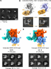
Fig. 1.
Fig. 1.. Cryo-EM data processing of HER2-TZB dataset showing the extended and compact HER2 conformations in complex with TZB.
(A) Representative 2D averages showing distinct secondary structure features of different views of the complex. Densities corresponding to the HER2 ECD or the Fab portion of TZB are labeled. Scale bar, 100 Å. (B) Top: Detail of two 3D classes representing two distinct conformations of the ECD in the HER2-TZB complex: extended (gray) and compact (purple). Dashed outline around the ECD head indicates the boundaries of the mask used for focused refinement in downstream steps. Density of TZB Fab is colored in light brown. For a more detailed workflow, refer to fig. S2. Bottom: 2D averages with particles (ptcls) filtered by angular assignment, such that only projections of “front views” were used in the average for each subset of particles. (C) Sharpened volume showing the extended conformation of HER2-TZB after focused refinement on the ECD head. (D) Sharpened volume of the compact HER2-TZB after increasing the number of particles of this conformation, followed by focused refinement on the ECD head. For clarity, density maps of the ECDs display subdomains in different colors. Subdomain I: From residues T23 to L215; subdomain II: T216 to C338; subdomain III: A339 to P500; subdomain IV: E501 to N629. Below each map, a collection of 2D averages of the ECD head after subtraction of the subdomain IV and TZB signal from each subset of particles, showing the average length of the ECD head (the measured distance is indicated with arrows). Scale bars, 50 Å.
Fig. 2.
Fig. 2.. Comparison of HER2 ECD models from this study with other HER2 models available in the PDB.
(A) RMSD of models of HER2 available in PDB. As reference, we used the Apo model obtained from the Apo dataset in the present study (Fig. 3A). Only distances between pairs of Cα atoms were considered for RMSD calculation. (B) Interdomain distance in angstrom between centroids of subdomains I and III measured on models of HER2 available in the PDB and compared with those of the present work. The different models are identified with their PDB accession numbers. Colors indicate the experimental technique used to obtain the structures.
Fig. 3.
Fig. 3.. Comparison of atomic models of the two conformations of the HER2-TZB complex.
(A) Overlay of backbones of atomic models of Apo and extHER2 ECDs. For RMSD, the Apo model was used as a reference. (B) Overlay of backbones of atomic models of extended and compact TZB-bound HER2 ECDs, aligned on subdomain III. Left: Front view. Right: Side view (subdomain II is hidden for clarity). For RMSD, either the Apo or the extHER2 model was used as a reference, as indicated by the subscript. (C) Comparison of individual subdomains of extHER2-TZB and compHER2-TZB. Insets show the subdomain being compared and the direction of view. Left: Subdomain I. Middle: Subdomain II. Right: Subdomain III. For RMSD, the extHER2-TZB model was used as a reference. (D and E). Cα-Cα distance between pairs of selected residues from subdomains I, II, and III at the crevice in the extended and compact models, respectively.
Fig. 4.
Fig. 4.. Superposition of the monomeric compact and extended conformations of the HER2 ECD-TZB complex on the cryo-EM HER2-HER3-NRG1β-TZB complex.
(A) Dimerization interface of several HER2 ECD models interacting with the HER3 dimerization arm. The specific HER2 model used in the figure is indicated in the text above it; in all cases, models were aligned on subdomain II, to visualize a hypothetical position of the HER3 dimerization arm in the dimerization arm-binding pocket. In orange, ribbon depiction of HER3 subdomain II. In dark blue, surface representation of the HER2 ECD head as it is in the model from the study of Diwanji et al. (30) (left) and after being substituted by the extHER2-TZB model (middle). In cyan, the HER2 model from (30) was substituted by the compHER2-TZB model (right) from this study. Residues with steric clashes are highlighted in red. (B) Detail of the dimerization arm-binding pockets from the insets in (A), with HER2 in ribbon depiction. In red, residues of HER2 involved in steric clashes. For clarity, only clashes between backbone atoms are displayed (dashed lines in light green).

References

    1. Yarden Y., Sliwkowski M. X., Untangling the ErbB signalling network. Nat. Rev. Mol. Cell Biol. 2, 127–137 (2001). - PubMed
    1. Olayioye M. A., Neve R. M., Lane H. A., Hynes N. E., The ErbB signaling network: Receptor heterodimerization in development and cancer. EMBO J. 19, 3159–3167 (2000). - PMC - PubMed
    1. Arkhipov A., Shan Y., Kim E. T., Dror R. O., Shaw D. E., Her2 activation mechanism reflects evolutionary preservation of asymmetric ectodomain dimers in the human EGFR family. eLife 2, e00708 (2013). - PMC - PubMed
    1. Jost C., Stüber J. C., Honegger A., Wu Y., Batyuk A., Plückthun A., Rigidity of the extracellular part of HER2: Evidence from engineering subdomain interfaces and shared-helix DARPin-DARPin fusions. Protein Sci. 26, 1796–1806 (2017). - PMC - PubMed
    1. Cho H. S., Mason K., Ramyar K. X., Stanley A. M., Gabelli S. B., Denney D. W. Jr., Leahy D. J., Structure of the extracellular region of HER2 alone and in complex with the Herceptin Fab. Nature 421, 756–760 (2003). - PubMed