

Results of arthroscopic microfracture treatment for traumatic and non-traumatic osteochondral lesions of the talus: a retrospective cohort study
- PMID: 40713601
- PMCID: PMC12297798
- DOI: 10.1186/s12891-025-08949-6
Results of arthroscopic microfracture treatment for traumatic and non-traumatic osteochondral lesions of the talus: a retrospective cohort study
Abstract
Background: This study is designed to assess the extent to which the outcomes of arthroscopic microfracture surgery for talus osteochondral lesions (OLTs)-whether of traumatic or atraumatic origin-are influenced by these underlying etiologic factors. Toward this end, it aims to optimise patient selection and treatment plans, thereby enabling the prediction of surgical prognosis.
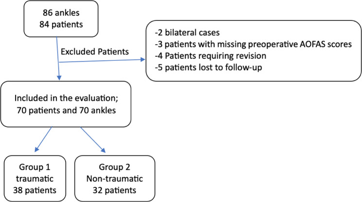
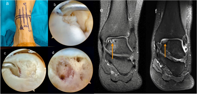
Methods: This retrospective study included 70 ankles from 70 patients with OLTs, who were treated with microfracture procedures using anterior ankle arthroscopy by orthopaedic surgeons at two different medical centres. Of these cases, 38 were of a traumatic origin (Group 1) and 32 were of a non-traumatic origin (Group 2). The inclusion criteria were adult patients with unilateral, detached and/or displaced lesions located in the medial central region of the talus. Preoperative and final follow-up American Orthopaedic Foot and Ankle Society (AOFAS) and Visual Analogue Scale (VAS) pain scores were compared within and between groups. The rate of return to baseline activity levels was also compared between the various groups. The potential influence of body mass index (BMI) on both etiology and surgical outcome was examined.
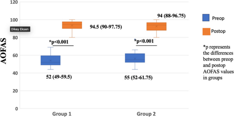
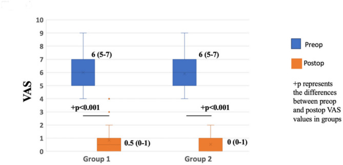
Results: The median follow-up period, as the interquartile range, for all patients was 39 months, ranging from 26 to 54.25 months. Both groups of patients showed significant improvement in their AOFAS and VAS scores postoperatively compared with their preoperative assessment (p < 0.001). Nevertheless, no statistically significant difference was found in the median AOFAS and VAS scores between Group 1 and Group 2 (p > 0.05). After the operation, 66 patients, representing 94.3%, successfully resumed their previous lifestyle, with no difference observed between the two groups (p = 0.392). In addition, the mean BMI in Group 2 was significantly higher than in Group 1 (p = 0.0035).
Conclusion: Arthroscopic microfracture treatment provides similar clinical outcomes in the case of non-traumatic and traumatic OLTs. A high BMI, however, has been recognized as a significant risk factor for the development of non-traumatic OLTs.
Keywords: Microfracture; Osteochondral lesion; Talus.
© 2025. The Author(s).
Conflict of interest statement
Declarations. Ethics approval and consent to participate: This study was approved by the Ethics Committee of Yeni Yüzyıl University (Approval No: 2025/04-1530). All participants in the study provided written informed consent before their involvement, in accordance with the principles outlined in the Declaration of Helsinki. Consent for publication: Not applicable. Competing interests: The authors declare no competing interests.
Figures
References
-
- Easley ME, Latt LD, Santangelo JR, Merian-Genast M, Nunley JA. Osteochondral lesions of the talus. J Am Acad Orthop Surg. 2010;18(10):616–30. - PubMed
-
- Van Bergen CJA, Kox LS, Maas M, Sierevelt IN, Kerkhoffs GMMJ, van Dijk CN. Arthroscopic treatment of osteochondral defects of the talus outcomes at eight to Twenty years of Follow-up 2013 by the journal of bone and joint surgery, incorporated. J Bone Joint Surg Am. 2013;95(6):519–25. - PubMed
-
- Griffith JF, Lau DT, Yeung DK, Wong MW. High-resolution MR imaging of Talar osteochondral lesions with new classification. Skeletal Radiol. 2012;41(4):387–99. - PubMed
Publication types
MeSH terms
LinkOut - more resources
Full Text Sources
Medical
