. 2025 Jul 26;15(1):19.
doi: 10.1186/s13395-025-00391-5.

Large MAF transcription factors reawaken evolutionarily dormant fast-glycolytic type IIb myofibers in human skeletal muscle

Affiliations

Large MAF transcription factors reawaken evolutionarily dormant fast-glycolytic type IIb myofibers in human skeletal muscle

Shunya Sadaki et al. Skelet Muscle. .

Abstract

Background: Small mammals such as mice rely on type IIb myofibers, which express the fast-contracting myosin heavy chain isoform Myh4, to achieve rapid movements. In contrast, larger mammals, including humans, have lost MYH4 expression. Thus, they favor slower-contracting myofiber types. However, the mechanisms underlying this evolutionary shift remain unclear. We recently identified the large Maf transcription factor family (Mafa, Mafb, and Maf) as key regulators of type IIb myofiber specification in mice. In this study, we investigate whether large MAFs play a conserved role in the induction of MYH4 expression and glycolytic metabolism in human and bovine skeletal muscle.

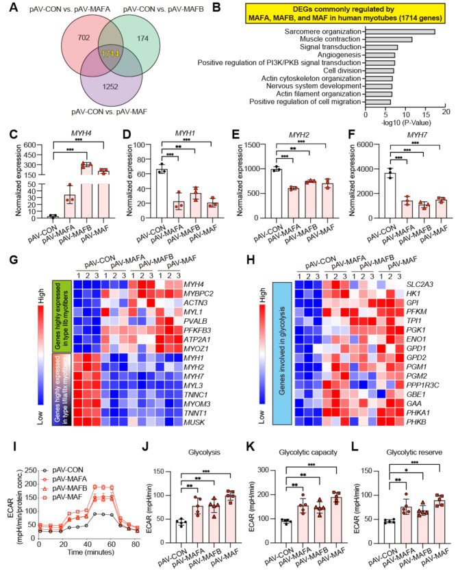
Methods: We performed adenovirus-mediated overexpression of large MAFs in iPSC-derived human myotubes and primary bovine myotubes. We subsequently quantified MYH4 expression using RT-qPCR, RNA sequencing (RNA-seq), and LC-MS/MS analysis. Glycolytic capacity was assessed using a flux analyzer and metabolic gene expression profiling. Additionally, RNA-seq analysis of human muscle biopsy samples was conducted to determine the correlations between large MAFs and the expression of MYH4 and other myosin genes, as well as their association with fast fiber composition and athletic training.

Results: Overexpression of large MAFs in human and bovine myotubes robustly induced MYH4 expression, with mRNA levels increasing by 100- to 1000-fold. LC-MS/MS analysis provided clear evidence of MYH4 protein expression in human myotubes, where it was previously undetectable. RNA-seq and flux analyzer data revealed that large MAFs significantly enhanced glycolytic capacity by upregulating the expression of key genes involved in glucose metabolism. Moreover, RNA-seq analysis of human muscle biopsy samples revealed a positive correlation between MAFA, MAF, and MYH4 expression. Furthermore, MAFA and MAF expression levels were elevated in power-trained individuals, accompanied by increased expression of MYH4 and other fast myosin genes.

Conclusions: Our findings establish large MAF transcription factors as key regulators of MYH4 expression and glycolytic metabolism in human skeletal muscle. This discovery provides novel insights into the evolutionary loss of type IIb myofibers in larger mammals and suggests potential strategies for enhancing muscle performance and mitigating fast-twitch fiber loss associated with aging and muscle degeneration.

Keywords: Aging; Human muscle; Large MAFs; MYH4; Myofiber type; Myosin heavy chain; Type IIb myofiber.


Conflict of interest statement

Declarations. Ethics approval and consent to participate: The Russian part of the study was approved by the Ethics Committee of the Federal Research and Clinical Center of Physical–Chemical Medicine of the Federal Medical and Biological Agency of Russia (protocol no. 2017/04), whereas the Finnish part of the study was approved by the Hospital District of Helsinki and Uusimaa (Database of Genotypes and Phenotypes Study Accession: phs001048.v2.p1). Written informed consent was obtained from all study participants. Consent for publication: Not applicable. Competing interests: TK and ST are inventors on a patent application related to this work (Patent Application No. 2022-083553). All other authors declare they have no competing interests.

Figures

Fig. 1
Fig. 1
MYH4 expression fluctuates in response to large MAF expression. (A) Correlation between the expression of large MAFs (MAFA, MAFB, and MAF) and MYH4 across multiple animal species. (B) Presence of MARE-like sequences in the promoter region of Myh4 across multiple animal species. (C) Visualization of MAFA, MAFB, and MAF expression using UMAP, as derived from single-nucleus RNA sequencing (snRNA-seq) of human hindlimb muscle samples from individuals aged 15–99 years (n = 27). Data sourced from Lai et al., Nature (2024). (D) Visualization of MYH4, MYH1, MYH2, and MYH7 expression using UMAP, as derived from snRNA-seq of human hindlimb muscle samples from individuals aged 15–99 years (n = 27). Data sourced from Lai et al., Nature (2024). (E) Average expression levels of MAFA, MAFB, MAF, MYH4, MYH1, MYH2, and MYH7 in myonuclei, expressed as the mean value for each individual in the respective age groups (young: 15–46 years, n = 8; old: 74–99 years, n = 19). P values were calculated using a two-sided Pearson correlation test (A) and an unpaired two-tailed t-test (E); *P < 0.05, **P < 0.01, ***P < 0.001, ****P < 0.0001, R; Pearson correlation coefficient
Fig. 2
Fig. 2
Large MAFs can specifically induce MYH4 mRNA expression in human iPSC-derived myotubes. (A) A schematic diagram illustrating adenovirus-mediated overexpression of MAFA, MAFB, and MAF in human iPSC-derived myotubes. After myogenic induction, iMuSCs were purified using flow cytometry and cultured in growth medium for expansion. After two days of exposure to differentiation medium, human myotubes were treated with adenoviral particles expressing MAFA_mCherry (pAV-MAFA), MAFB_mCherry (pAV-MAFB), or MAF_mCherry (pAV-MAF). mCherry-only was used as the control (pAV-CON). Created in BioRender. Ryo, F. (2025) https://BioRender.com/r49s132. (B) Immunostaining of human myotubes transduced with pAV-CON, pAV-MAFA, pAV-MAFB, or pAV-MAF using antibodies against alpha-actinin (α-ACTININ, green) and mCherry (red). Nuclei were stained with DAPI (blue). (C) Quantification of infection efficiency for each adenoviral vector. The proportion of mCherry-positive cells within α-ACTININ-positive myotubes was calculated. In total, 91–105 myotubes were analyzed per experiment (n = 4). (D) Quantification of myotube width shown in panel (B). In total, 157–283 myotubes were analyzed per experiment (n = 4). (E) Relative mRNA expression levels of MAFA, MAFB, and MAF, as determined using RT-qPCR in human myotubes 4 days after adenovirus transduction (n = 3/group). (F) Detection of MYH4 mRNA in human myotubes transduced with pAV-CON, pAV-MAFA, pAV-MAFB, or pAV-MAF using RNAscope. Myotubes were visualized with an antibody against α-ACTININ (white) and mCherry (red) after hybridization. Nuclei were stained with DAPI (blue). Scale bar: 50 μm. (G-I) Relative mRNA expression levels of myosin heavy chain genes (MYH1, MYH2, MYH4, and MYH7), as determined using RT-qPCR in human myotubes 4 days after transduction with adenoviral vectors expressing MAFA (G), MAFB (H), or MAF (I). mCherry-only was used as the control (pAV-CON, n = 3/group). P values were calculated using Tukey’s test (C, D) and Student’s t-test (E, G-I); *P < 0.05, **P < 0.01, ***P < 0.001
Fig. 3
Fig. 3
MYH4 mRNA induced by large MAFs is translated into protein in human myotubes. (A) A schematic diagram illustrating the protocol for isolating active ribosomes to detect MYH4 mRNA translation in human myotubes following adenoviral overexpression of MAFA, MAFB, or MAF. Created in BioRender. Ryo, F. (2025) https://BioRender.com/g05y857. (B) The amplification curve of MYH4 mRNA bound to active ribosomes in human myotubes, as determined using RT-qPCR. (C) Gel electrophoresis of qPCR products corresponding to MYH4 and TBP mRNAs bound to active ribosomes, as analyzed in panel (B). (D) A schematic diagram illustrating the protocol for the detection of MYH4 using LC-MS/MS in human myotubes following adenoviral overexpression of MAFA, MAFB, or MAF. Created in BioRender. Ryo, F. (2025) https://BioRender.com/f96f475. (E-G) MAFA (E), MAFB (F), and MAF (G) expression in human myotubes following adenoviral overexpression of MAFA, MAFB, or MAF, as determined via LC-MS/MS. mCherry-only was used as the control (pAV-CON) (n = 3/group). N.D.; not detected. (H) The number of MYH4-specific peptides detected in human myotubes following adenoviral overexpression of MAFA, MAFB, or MAF, as determined using LC-MS/MS. mCherry-only was used as the control (pAV-CON, n = 3/group). N.D.; not detected. (I) Percent peptide coverage of MYH4 in LC-MS/MS analysis. Coverage is expressed as the percentage of the amino acid sequences detected via LC-MS/MS analysis
Fig. 4
Fig. 4
Large MAFs specifically upregulate the expression of type IIb myofiber-associated and glycolytic genes, consequently enhancing glycolysis in human myotubes. (A) Venn diagrams showing the differentially expressed genes (DEGs) identified in comparisons between pAV-CON and pAV-MAFA (3,685 genes), pAV-CON and pAV-MAFB (2,289 genes), and pAV-CON and pAV-MAF (4,446 genes). In total, 1,714 DEGs were commonly altered by the overexpression of large MAFs. (B) Gene ontology (GO) analysis of the 1,714 commonly altered genes identified in panel (A). (C-F) RNA-seq analysis of MYH4, MYH1, MYH2, and MYH7 expression in human myotubes transduced with pAV-CON, pAV-MAFA, pAV-MAFB, or pAV-MAF (n = 3/group). (G) A heatmap visualizing the expression of type IIb myofiber-associated genes and other myofiber type-associated genes (n = 3/group). (H) A heatmap visualizing the expression of genes involved in glycolysis (n = 3/group). (I) Extracellular acidification rate (ECAR) in human myotubes transduced with pAV-CON, pAV-MAFA, pAV-MAFB, or pAV-MAF, as measured using the Seahorse XFe24 Analyzer (n = 5/group). (J-L) Glycolysis, glycolytic capacity, and glycolytic reserve calculated from the ECAR data shown in panel (I). P values were calculated using Tukey’s test (C-F, J-L); *P < 0.05, **P < 0.01, ***P < 0.001, **** P < 0.0001
Fig. 5
Fig. 5
Analysis of human skeletal muscle biopsy samples reveals a positive correlation between MYH4 and large MAF expression, highlighting the potential functionality of large MAFs in human skeletal muscle tissue. (A-B) Muscle biopsy samples were obtained from the vastus lateralis of 291 individuals in Finland, comprising 166 men and 125 women aged 20–79 years (mean age: 59.9 years). (A) Scatter plots showing the percentage of fast-twitch myofibers (y-axis) against the mRNA expression levels of MAFA, MAFB, MAF, or total large MAFs (MAFA + MAFB + MAF) (x-axis) for each participant, as determined using RNA-seq analysis. (B) Scatter plots showing the mRNA expression levels of MYH4, MYH1, MYH2, or MYH7 (y-axis) against the total mRNA expression levels of large MAFs (MAFA + MAFB + MAF) (x-axis) for all 291 participants. (C-D) Muscle biopsy samples were obtained from the vastus lateralis of 24 men in Russia. The ages of the participants ranged from 20 to 45 years (mean age: 32.7 years). (C) Scatter plots showing the percentage of fast-twitch myofibers (y-axis) against the mRNA expression levels of MAFA, MAFB, MAF, or total large MAFs (MAFA + MAFB + MAF) (x-axis) for each participant, as determined through immunohistochemical and RNA-seq analyses. (D) Scatter plots showing the mRNA expression levels of MYH4, MYH1, MYH2, or MYH7 (y-axis) against the total mRNA expression levels of large MAFs (MAFA + MAFB + MAF) (x-axis) for all 24 participants. (E) RNA-seq analysis of MAFA, MAFB, MAF, MYH4, MYH1, MYH2, and MYH7 expression in Russian muscle biopsy samples, categorized by training type: endurance training (n = 14) and power training (n = 10). P values were calculated using a two-sided Pearson correlation test (A-D) and Student’s t-test (E); *P < 0.05, **P < 0.01, ***P < 0.001, R; Pearson correlation coefficient. Outliers were identified using Grubbs’ test (Alpha = 0.05); three participants with outlying MYH4 expression levels were excluded from the analysis (D-E)

References

    1. Ciciliot S, Rossi AC, Dyar KA, Blaauw B, Schiaffino S. Muscle type and fiber type specificity in muscle wasting. Int J Biochem Cell Biol. 2013;45:2191–9. - PubMed
    1. Schiaffino S, Reggiani C. Fiber types in mammalian skeletal muscles. Physiol Rev. 2011;91:1447–531. - PubMed
    1. Bottinelli R, Canepari M, Pellegrino MA, Reggiani C. Force-velocity properties of human skeletal muscle fibres: myosin heavy chain isoform and temperature dependence. J Physiol. 1996;495:573–86. - PMC - PubMed
    1. Schiaffino S, Chemello F, Reggiani C. The diversity of skeletal muscle Fiber types. Cold Spring Harb Perspect Biol. 2024;a041477. - PubMed
    1. Albers PH, Pedersen AJT, Birk JB, Kristensen DE, Vind BF, Baba O, et al. Human muscle fiber type-specific insulin signaling: impact of obesity and type 2 diabetes. Diabetes. 2015;64:485–97. - PubMed

Substances

LinkOut - more resources