Hounsfield Unit characterization and dose calculation on a C-arm linac with novel on-board cone-beam computed tomography feature and advanced reconstruction algorithms
- PMID: 40714930
- PMCID: PMC12301083
- DOI: 10.1002/acm2.70145
Hounsfield Unit characterization and dose calculation on a C-arm linac with novel on-board cone-beam computed tomography feature and advanced reconstruction algorithms
Abstract
Purpose: Current cone-beam computed tomography (CBCT) on-board c-arm linear accelerators (linacs) lack CT number precision sufficient for dose calculation due to increased scatter from the cone geometry. This investigation evaluated CT number and dose calculation accuracy in-phantom on a novel on-board CBCT unit with potential for improved dose calculation accuracy.
Methods: Eight head and eight body configurations of an electron density phantom using 16 materials were acquired with a clinical CT-simulator(CT-sim) and novel on-board CBCT imager. CBCT projection data was reconstructed using conventional Feldkamp-Davis-Kress(FDK) and patient scatter-corrected iterative Acuros CTS with metal artifact reduction (Acuros-CTS-iCBCT-MAR) to create a robust CT number to physical density curve. CT-sim and CBCT images of anthropomorphic head and thorax phantoms were acquired, and three treatment plans were generated per phantom. All CBCT images were registered to the CT-sim of the phantoms, and the dose was recalculated on the CBCT images. 3D gamma analysis was performed (10% dose threshold, local, 1% dose difference, 1 or 2 mm distance to agreement), and dose-volume histogram (DVH) metrics were reported for target coverage and organ sparing.
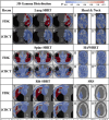
Results: CT numbers for materials ≤1.08 g/cc showed high agreement between CT-sim and CBCT acquisitions. CT number precision improved with Acuros-CTS-iCBCT-MAR compared to FDK in all materials. High agreement between CT-sim and CBCT reconstructed with Acuros-CTS-iCBCT-MAR was observed in 3D gamma analysis showing 93.8% voxels passing in the worst case for a spine stereotactic body radiotherapy (SBRT) plan at 1%/1 mm. Maximum deviation in target coverage was -3.3% PTV-D98% in the lung SBRT plan among the novel reconstructed CBCT images. Plan comparison using FDK reconstructions yielded similar or worse agreement in 3D gamma analysis, with 69.7% voxels passing in the worst case for a spine SBRT treatment plan. Maximum target coverage deviation was -11.7% PTV-D98% among the FDK-reconstructed CBCT.
Conclusions: Novel CBCT solutions on c-arm linacs show promise for enabling direct-to-unit or offline adaptive dose calculation, potentially increasing versatility and efficiency of patient-centered care.
Keywords: Hounsfield Unit characterization; dose calculation; fast cone‐beam CT; inhomogeneity correction.
© 2025 The Author(s). Journal of Applied Clinical Medical Physics published by Wiley Periodicals LLC on behalf of American Association of Physicists in Medicine.
Conflict of interest statement
Kenneth Gregg, Theodore Arsenault, Atefeh Rezaei, and Rojano Kashani report no conflicts of interest related to this project. Alex Price has received funding from Radformation for giving research presentations. Lauren Henke has received funding from Varian Medical Systems for consulting and giving research presentations and is on the Elekta medical advisory board. University Hospitals was funded with an industry grant by Varian Medical Systems.
Figures


References
-
- Papanikolaou N, Battista JJ, Boyer AL, et al. Tissue Inhomogeneity Corrections for Megavoltage Photon Beams (AAPM Report No. 85, 2004). doi: 10.37206/86 - DOI
MeSH terms
LinkOut - more resources
Full Text Sources
Research Materials

