. 2025 Jul 26;16(1):6903.
doi: 10.1038/s41467-025-62031-2.

Melbournevirus encodes a shorter H2B-H2A doublet histone variant that forms structurally distinct nucleosome structures

Affiliations

Melbournevirus encodes a shorter H2B-H2A doublet histone variant that forms structurally distinct nucleosome structures

Alejandro Villalta et al. Nat Commun. .

Abstract

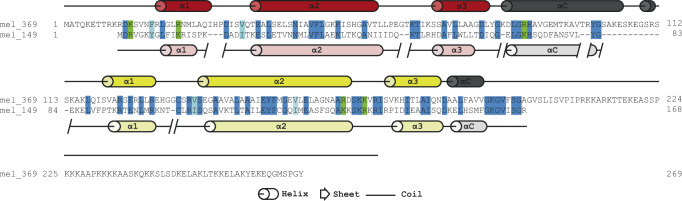
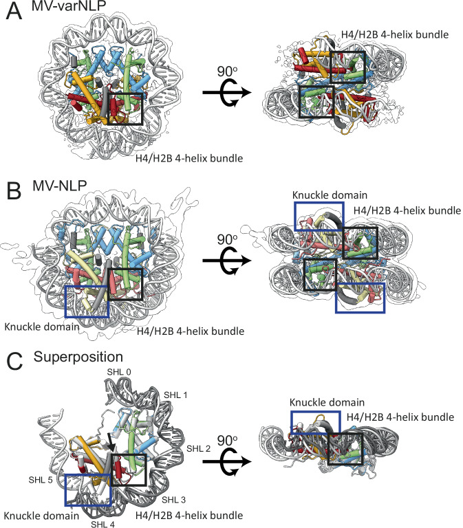
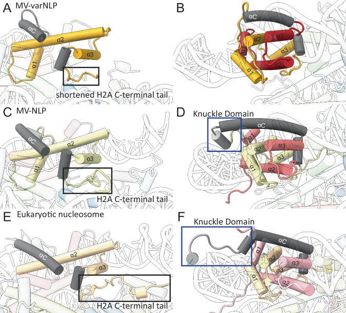
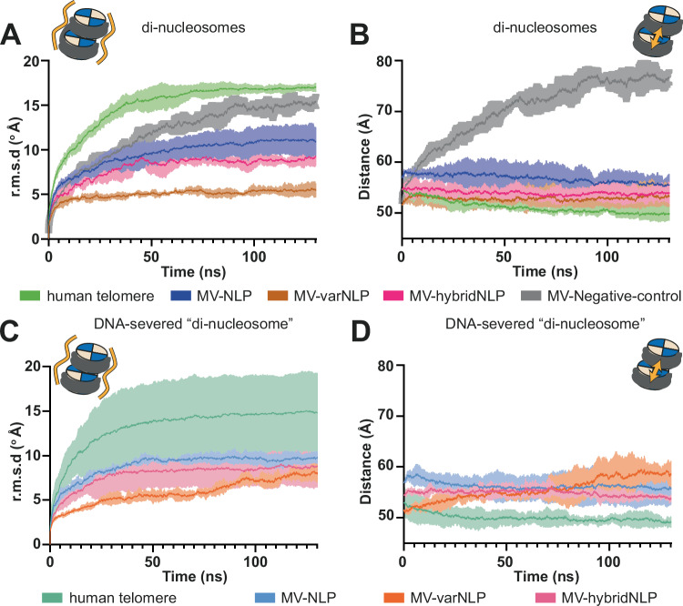
Unique among viruses, some giant viruses utilize histones to organize their genomes into nucleosomes. Melbournevirus encodes a distinct H2B-H2A histone doublet variant in addition to the canonical H4-H3 and H2B-H2A doublets. This viral histone variant has a truncated H2B portion and its amino acid sequence deviates from that of the main viral H2B-H2A throughout the entire coding region. It is less abundant than the main H2B-H2A doublet, is likely essential for melbournevirus fitness, and is conserved in all Marseilleviridae. The cryo-EM structure of a nucleosome-like particle reconstituted with this H2B-H2A variant and viral H4-H3 reveals that only 90 base pairs of DNA are stably bound, significantly less than in eukaryotic nucleosomes and viral nucleosomes made with the main fused viral histone doublets. The reduced ability to bind DNA can be attributed to structural differences between variant and main H2B-H2A. Variant melbournevirus nucleosomes are less stable, possibly aiding rapid genome unpacking to initiate gene expression. Our results highlight the remarkable propensity of giant viruses to appropriate the utility of histones for their specialized purposes.


Conflict of interest statement

Competing interests: The authors declare no competing interests.

Figures

Fig. 1
Fig. 1. Melbournevirus encodes an H2B-H2A histone variant.
The amino acid sequence of MV-H2B-H2A (mel_369, secondary structure elements taken from pdb 7N8N shown in dark red for H2B, and yellow for H2A) and MV-varH2B-H2A (mel_149, predicted secondary structure elements shown in light red and light yellow for H2B and H2A, respectively). Dark blue highlighted amino acids (aa) are conserved between the two proteins; similar hydrophobic amino acids are shown in light blue (V/I or F/W/Y), and conserved positively charged aa (R/K) are highlighted in green.
Fig. 2
Fig. 2. mel_149 deletion strongly impacts melbournevirus fitness.
A Schematic representation of the strategy used to generate the variant H2B-H2A deleted virus. The selection cassette was introduced by homologous recombination. The location of the primers used for genotyping (a, b, and c) are indicated. B Variant H2B-H2A deleted viruses were generated, selected, and cloning was attempted as indicated by the timeline. After passage 4 in presence of selection (nourseothricin), since we were unable to obtain pure variant H2B-H2A deleted viruses; viruses were split and used to infect non-complemented amoebae (P5*) or trans-complementing amoebae (P5) in absence of selection. Trans-complementation refers to the restoration of the function of a deleted or defective gene in a viral genome through the expression of the same gene from an independent genetic element, such as the host genome or a plasmid. Subsequent passages were performed and genotyped to assess the fitness associated with gene knockout in competition with wild type viruses. C PCR amplifications of mel_149 at three cellular passages of A. castellanii, complemented with mel_149 or wildtype. Genotyping was performed using the primers indicated in (A) and the experiment was performed as shown in (B). This image is representative of 2 independent experiments.
Fig. 3
Fig. 3. MV-varNLP is structurally similar to MV-NLP.
Experimental electron density and model of (A) MV-varNLP, and (B) MV-NLP (PDB: 7N8N). The histone fold regions of H2B are shown in red, H2A in yellow, H4 in green, and H3 in blue, with helices not belonging to the histone fold in grey. C Superposition of MV-varNLP with MV-NLP. Here, only half of the nucleosome is shown for clarity. Superhelix locations (SHL) are numbered as previously defined. The four-helix bundle region tethering H4 to H2B, and the “knuckle domain” that binds DNA in MV-NLP are indicated by black and blue boxes, respectively. The arrow corresponds to the H2B α3-αC helix interface.
Fig. 4
Fig. 4. MV-varNLP and MV-NLP differ in their H2B portion and in the extent of DNA binding.
Cartoon representation of parts of H2B and the H2A C-terminal tail and knuckle domain of (A, B) MV-varNLP, (C, D) MV-NLP (7N8N), and (E, F) the eukaryotic nucleosome (1AOI). Models are represented in transparent colors with solid colors at regions of interest (H2A, yellow; H2B, red; knuckle domain, gray).
Fig. 5
Fig. 5. MV-NLP can form nucleosome stacks in silico.
Two parameters were measured to assess the stability of di-nucleosome stacks: The mean variation of (A, C) the root mean square deviation (r.m.s.d) of the overall structure compared to the start of the simulation, and (B, D) the distances between the center of gravity of the two nucleosomes over the simulation time are plotted for the human telomeric di-nucleosome (green), MV-NLP (blue), MV-varNLP (brown), MV-hybridNLP (purple) and a control for MV-varNLP wherein inter-nucleosomal protein-protein contacts were mutated in silico to Glu (grey). C, D show a simulation of di-nucleosomes where 10 bp at the DNA linker portion connecting both nucleosomes were removed to sever the connection between the two particles. The larger r.m.s.d variations at the beginning of the simulation for the human telomeric di-nucleosome are due to the long tails that collapse onto the DNA, as previously observed for eukaryotic mono-nucleosomes. The lighter area around the mean curve corresponds to the standard variation around the mean curve, from three replicates. Individual tracks are found in Supplementary Fig. 7.

Similar articles

Cited by

References

    1. Luger, K., Mäder, A. W., Richmond, R. K., Sargent, D. F. & Richmond, T. J. Crystal structure of the nucleosome core particle at 2.8 Å resolution. Nature389, 251–260 (1997). - PubMed
    1. Stevens, K. M. & Warnecke, T. Histone variants in archaea – An undiscovered country. Semin. Cell Dev. Biol.135, 50–58 (2023). - PubMed
    1. Grau-Bové, X. et al. A phylogenetic and proteomic reconstruction of eukaryotic chromatin evolution. Nat. Ecol. Evol.6, 1007–1023 (2022). - PMC - PubMed
    1. Laursen, S. P., Bowerman, S. & Luger, K. Archaea: the final frontier of chromatin. J. Mol. Biol.433, 166791 (2021). - PMC - PubMed
    1. Mattiroli, F. et al. Structure of histone-based chromatin in Archaea. Science357, 609–612 (2017). - PMC - PubMed

LinkOut - more resources