. 2025 Jul 25;16(1):6843.
doi: 10.1038/s41467-025-62118-w.

125Te and 57Fe nuclear resonance vibrational spectroscopic characterization of intermediate spin state mixed-valent dimers

Affiliations

125Te and 57Fe nuclear resonance vibrational spectroscopic characterization of intermediate spin state mixed-valent dimers

Aleksa Radović et al. Nat Commun. .

Abstract

Iron-sulfur clusters fulfill numerous roles throughout biology. The reduced [2Fe-2S]+ cluster offers unique electronic and magnetic properties due to its mixed-valent nature and can serve as an essential model for understanding electron transfer, electron delocalization, and accessible spin states not only in mixed-valent dimers, but potentially larger iron sulfur clusters. Recently a series of mixed-valent diiron dichalcogenide complexes [L2Fe2Q2]- (Q = S (1), Se (2), Te (3), L = 2,6-diisopropylphenyl β-diketiminate ligand) were synthesized and characterized, where complex 1 showed a typical S = 1/2 spin state, while complexes 2 and 3 exhibited intermediate S = 3/2 spin states, potentially enabled by the minimization of vibronic coupling. Here we studied the vibrational dynamics of the Fe and Te centers in these complexes using 57Fe and 125Te nuclear resonance vibrational spectroscopy (NRVS), coupled with DFT calculations. The findings suggest that heavy character of larger chalcogen atoms results in decreased vibronic coupling. The observation of an intermediate spin state is shown to be unattainable for lighter Fe2Q2 cores. This highlights the crucial role of vibronic coupling in modulating the electronic structure of mixed-valence systems and should enhance understanding of the electronic structure in more complex biological Fe-S clusters.


Conflict of interest statement

Competing interests: The authors declare no competing interests.

Figures

Fig. 1
Fig. 1. Structures of studied complexes and example of potential energy surfaces.
a Structures of previously synthesized mixed-valent diiron dichalcogenide clusters. b Energy levels determined by Eq. 1, as a function of B/|J| ratio, for antiferromagnetically coupled systems (J < 0). c Representation of PKS vibration for studied complexes. d, e Energy levels determined by Eq. 2 in the PKS coordinate for d S = 1/2 localized [Fe2S2]+ complex and e S = 9/2 delocalized [Fe2(OH)3(tmtacn)2]2+,.
Fig. 2
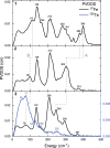
Fig. 2. Experimental 57Fe and 125Te nuclear resonance vibrational spectroscopy (NRVS) partial vibrational density of states (PVDOS).
57Fe (black line) and 125Te (blue line) NRVS PVDOS spectra of complexes 13. Gray dashed lines depict the shifting of the Fe-Q stretching (A) and lower energy core (B) vibrations.
Fig. 3
Fig. 3. Calculated 57Fe NRVS and PKS vibrational modes.
a Comparison of experimental (black line) and calculated (red line) 57Fe NRVS spectra of complexes 13. Individual transitions are depicted by red vertical lines. b 57Fe PVDOS along x (red line), y (blue line), and z (brown line) axes, for complexes 13. The x axis is defined along Fe-Fe bond, and z axis is perpendicular to the [Fe2Q2]+ core. c Representation of calculated PKS normal modes for complexes 13. For clarity, only the [Fe2Q2]+ cores and coordinating N atoms are shown.
Fig. 4
Fig. 4. Comparison of 57Fe and 125Te NRVS and selected vibrational modes.
a Comparison of 125Te (top) and 57Fe (bottom) NRVS spectra of complex 3. Experimental spectra are depicted by black lines, calculated spectra by blue (125Te) and red (57Fe) lines, with selected individual transitions depicted by blue and red vertical lines. b Representations of selected calculated normal modes of complex 3. For clarity, only the [Fe2Te2]+ core and coordinating N atoms are shown. Magenta colored rectangles represent regions containing normal modes with significant Te displacements (a) and corresponding normal modes (b). Orange colored rectangles represent regions containing normal modes with significant Fe displacements (a) and corresponding normal modes (b).
Fig. 5
Fig. 5. Potential energy surfaces and influence of vibronic coupling.
a Ground and excited spin states in the PKS coordinate for the complexes 13. b Spin states in the PKS coordinate for the complex 1 with a decreased amount of vibronic coupling. c Spin states in the PKS coordinate for the complex 3 with an increased amount of vibronic coupling.

Similar articles

References

    1. Beinert, H., Holm, R. H. & Munck, E. Iron-sulfur clusters: nature’s modular, multipurpose structures. Science277, 653–659 (1997). - PubMed
    1. Solomon, E. I., Xie, X. J. & Dey, A. Mixed valent sites in biological electron transfer. Chem. Soc. Rev.37, 623–638 (2008). - PubMed
    1. Albers, A. et al. The complete characterization of a reduced biomimetic [2Fe-2S] cluster. Angew. Chem. Int. Edit. 50, 9191–9194 (2011). - PubMed
    1. Saouma, C. T., Kaminsky, W. & Mayer, J. M. Protonation and concerted proton-electron transfer reactivity of a bis-benzimidazolate ligated [2Fe-2S] model for rieske clusters. J. Am. Chem. Soc.134, 7293–7296 (2012). - PMC - PubMed
    1. Albers, A., Bayer, T., Demeshko, S., Dechert, S. & Meyer, F. A Functional model for the Rieske center: full characterization of a biomimetic N-Ligated [2Fe-2S] cluster in different protonation states. Chem. Eur. J. 19, 10101–10106 (2013). - PubMed

LinkOut - more resources