. 2025 Jul 26;15(1):27176.
doi: 10.1038/s41598-025-07299-6.

In vivo efficacy of NRL knockdown with cell-penetrating siRNA in retinal degeneration

Affiliations

In vivo efficacy of NRL knockdown with cell-penetrating siRNA in retinal degeneration

Hyungwoo Lee et al. Sci Rep. .

Abstract

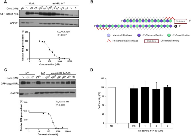
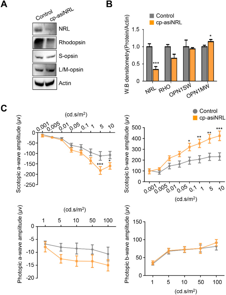
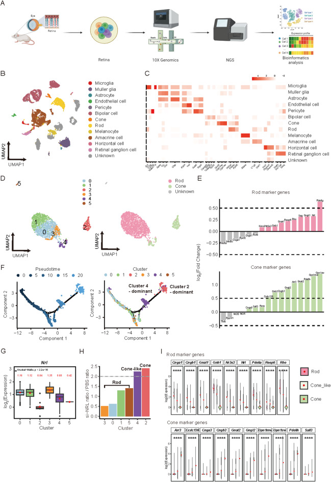
Retinal degenerative diseases, such as retinitis pigmentosa (RP) and age‑related macular degeneration (AMD), lead to progressive vision loss through photoreceptor degeneration; RP begins with the gradual loss of peripheral rods, whereas AMD causes central‑vision loss mainly because macular cones and parafoveal rods degenerate. The neural retina leucine zipper (NRL) directs rod photoreceptor differentiation, and its disruption has been linked to upregulated cone-specific markers in rods. This study investigates the therapeutic potential of a cell-penetrating asymmetric small interfering RNA targeting NRL (cp-asiNRL) to induce rod-to-cone conversion and mitigate retinal degeneration. cp-asiNRL was administered intravitreally to C57BL/6J wild-type (WT), neovascular AMD (nAMD), and RP (RhoP23H/+) mouse models. Subsequent analyses included cone marker expression levels and electroretinographic evaluations, and single-cell RNA sequencing. Administration of cp-asiNRL suppressed NRL expression, increased cone marker expression, and improved retinal function in both WT and nAMD models. In RP mice, cone marker expression was also elevated, although functional improvements were comparatively modest, likely reflecting the advanced disease stage. Single-cell RNA sequencing revealed a rod-to-cone-like transdifferentiation, suggesting that cp-asiNRL-mediated NRL knockdown partially preserved photoreceptor integrity. cp-asiNRL-mediated NRL silencing shows considerable promise as a therapeutic intervention for retinal degenerative conditions. By promoting rod-to-cone transdifferentiation and supporting photoreceptor survival, this approach may offer a novel strategy for vision preservation.


Conflict of interest statement

Competing interests: H.J., T.H., M.Y., S.P., S.W.H., and D.K.L. are employees of OliX Pharmaceuticals Inc. H.C. serves as a consultant for OliX Pharmaceuticals Inc. A related patent application has been filed: PCT/KR2021/018460. The remaining authors - H.L., J.C., H.K.L., C.S., C.P., S.Y.L., J.L., and S.L. - declare no competing interests.

Figures

Fig. 1
Fig. 1
Molecular design and selection of cell-penetrating asymmetric small interfering RNA targeting NRL (cp-asiNRL). (A) Half-maximal inhibitory concentration (IC50) of the potent asiNRL #47. HeLa cells overexpressing human NRL were incubated with various concentrations ranging from 0.01 to 3 nM asiNRL #47 after transfection for 48 hours, followed by western blotting analysis, and the bands were normalized to those of GAPDH. (B) To confer cell-penetrating ability to asiRNAs, chemical modifications were introduced into the sugar-phosphate backbone, and a cholesterol moiety was conjugated at the 3’ end of the sense strand. (C) IC50 of the comparative cp-asiNRL #47 − 19. The A549 cell line constitutively overexpressing GFP-tagged NRL (A549/NRL) was incubated in the range of 30 to 3000 nM for 72 h by passive uptake, followed by western blotting analysis, and the bands were normalized to those of GAPDH. (D) Cell viability of cells treated with cp-asiNRL #47 − 19. The cells were incubated for 24 h with various concentrations of cp-asiNRL #47 − 19 (0.5, 1, 3, and 5 µM), followed by the MTT assay. The data are shown as the mean ± SD of 3 independent experiments. NT; non-treated.
Fig. 2
Fig. 2
cp-asiNRL knockdown efficacy in WT mice. (A) Representative western blot images. (B) Relative densitometric bar graphs (n = 4 per group). (C) Immunohistochemistry images dorsal retinal sections showing cone and rod markers in WT mice. (D) Quantification of the number of cells displaying positive fluorescence for S-opsin and L/M-opsin in control and cp-asiNRL-treated retinas. Data are presented as mean ± SD (n = 3 per group). (E) Electroretinogram under scotopic and photopic conditions. Note that the scotopic a‑wave shows significance only at intensities ≥ 1 cd·s/m⁻², reflecting cone contribution after rod saturation. (n = 4 per group). *p < 0.05.
Fig. 3
Fig. 3
Cp-asiNRL knockdown efficacy in the CNV mouse model. (A) Representative western blot images. (B) Relative densitometric bar graphs (n = 3 per group). (C) Electroretinogram under scotopic and photopic conditions (n = 7 per group). *p < 0.05.
Fig. 4
Fig. 4
cp-asiNRL knockdown efficacy in RhoP23H/+ transgenic mice. (A) Representative western blot images. (B) Relative densitometric bar graphs (n = 4 per group). (C) Immunohistochemistry images showing cone- and rod-specific markers in the dorsal retina. (D) Quantification of the number of cells displaying positive fluorescence for S-opsin and L/M-opsin in control and cp-asiNRL-treated retinas. Data are presented as mean ± SD (n = 3 per group). (E) Representative images of apoptosis in the outer retinal layer were detected using a TUNEL assay. The blue color denotes all nuclei, and the green color denotes DNA-damaged nuclei. The graph shows a significantly decreased ratio of apoptotic nuclei in the cp-asiNRL-treated group compared to the control group. (F) Electroretinogram under scotopic and photopic conditions (n = 5 per group). *p < 0.05.
Fig. 5
Fig. 5
scRNA-seq data analysis. (A) Workflow of the scRNA-seq analysis. (B) UMAP of 13 cell types detected in retinal cells. (C) Heatmap of marker gene expression values in 13 cell types. (D) UMAP of photoreceptor cells. (E) Bar plots of log2(fold change) of marker genes in rod and cone photoreceptors; the black dashed line indicates a log2(fold change) of 0.5, the significance threshold, compared to the cp-asiNRL and PBS groups. (F) Trajectory analysis of photoreceptor cells using Monocle2. (G) The expression levels of the Nrl gene in 6 subclusters. (H) The ratio of the cp-asiNRL ratio to the PBS ratio to the cell count in 6 clusters; the gray the dashed line denotes the 2-fold significance cutoff. (I) Violin plot of marker genes in three types of cells, namely, rod, cone-like, and cone cells.

References

    1. Rim, T. H., Park, H. W., Kim, D. W. & Chung, E. J. Four-year nationwide incidence of retinitis pigmentosa in South Korea: a population-based retrospective study from 2011 to 2014. BMJ Open.7, e015531. 10.1136/bmjopen-2016-015531 (2017). - PMC - PubMed
    1. Li, J. Q. et al. Prevalence and incidence of age-related macular degeneration in Europe: a systematic review and meta-analysis. Br. J. Ophthalmol.104, 1077–1084 (2020). - PubMed
    1. Teo, C. L. et al. Prevalence of retinitis pigmentosa in singapore: the Singapore Epidemiology of Eye Diseases Study. Acta Ophthalmol.99, e134–e135. 10.1111/aos.14483 (2021). - PubMed
    1. de Cabral, T. A., Daich Varela, M., Georgiou, M. & Michaelides, M. Treatments for dry age-related macular degeneration: therapeutic avenues, clinical trials and future directions. Br. J. Ophthalmol.31845210.1136/bjophthalmol-2020-318452 (2021). - PMC - PubMed
    1. Schaffrath, K. et al. One-Year safety and performance assessment of the Argus II retinal prosthesis: a postapproval study. JAMA Ophthalmol.137, 896–902 (2019). - PMC - PubMed

MeSH terms

LinkOut - more resources