. 2025 Jul 25;15(1):27106.
doi: 10.1038/s41598-025-12926-3.

Regulation of RPE65 expression in human retinal pigment epithelium cells

Affiliations

Regulation of RPE65 expression in human retinal pigment epithelium cells

Olga A Postnikova et al. Sci Rep. .

Abstract

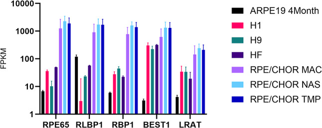
The visual cycle is an important pathway in the retinal pigment epithelium (RPE) which regenerates 11-cis retinal chromophore for the retinal photoreceptors. The central enzyme in the visual cycle is RPE65 retinol isomerase. Expression of RPE65 mRNA and protein levels are significantly lower in RPE cell culture models when compared to native RPE. This limits the use of these models to study the visual cycle. To determine the main drivers of RPE65 regulation we compared the transcriptional profiles of native and cell culture models of RPE with various levels of RPE65 expression. We also compared the levels of RPE65 expression between ARPE-19 cells grown in media supplemented with 1 mM pyruvate (PYR) or 10 mM nicotinamide (NAM). In addition, we performed experiments directed at transcriptional and translational regulation of RPE65. We show that RPE65 mRNA and protein expression is significantly higher in NAM media grown cells than PYR cells. Transfection of cells with a variety of different vectors containing RPE65 ORFs with different promoters, codon optimization, IRES, 3' UTRs, suggest that translational effects are less important than transcriptional status. Importantly, we found that feeding with rod outer segments (ROS) decreases RPE65 expression in NAM grown cells, suggesting that certain primary functions of the RPE (here, visual cycle and phagocytosis) are not positively linked. Analysis of differentially regulated microRNAs (miRs) provides a basis for this downregulation. It appears that the regulation of RPE65 expression in ARPE-19 cells, in particular, is multifactorial, involving primarily metabolic and transcriptional status of the cells, with translation of RPE65 mRNA playing a smaller role.

Keywords: MicroRNAs; Nicotinamide; Pyruvate; RPE65; Retina; Retinal pigment epithelium; Ribosome; Transcription; Translation.


Conflict of interest statement

Declarations. Competing interests: The authors declare no competing interests.

Figures

Fig. 1
Fig. 1
FPKM normalized expression levels of visual cycle genes from RNA-seq data. RNA-seq data was obtained from: ARPE-19 4 Month, ARPE-19 cells differentiated for 4 months under PYR protocol; H1, RPE differentiated from H1 ESC; H9, RPE differentiated from H9 ESC; HF, Human Fetal RPE cell lines; RPE/CHOR, RPE from macular (MAC), nasal (NAS), and temporal (TMP) regions of human RPE/choroid, respectively. Data was collected from (10, 15, 16).
Fig. 2
Fig. 2
Detection of RPE65 protein and mRNA expression in ARPE-19 cultured in NAM media. A. Detection of RPE65 protein with rabbit monoclonal RPE65-[EPR7024(N)]-C-terminal antibody (Abcam, Cat. No. ab175936). Bovine RPE microsomes were used as a positive control. Blots were visualized using a LI-COR Odyssey CLx Imaging System. B. qPCR quantification of RPE65 mRNA in ARPE-19 cells differentiated under NAM or PYR protocol.
Fig. 3
Fig. 3
Overrepresented pathways in genes upregulated in NAM grown ARPE-19 cells compared to those grown in PYR. KEGG 2019 pathways overrepresented in ARPE-19 cells grown in NAM protocol compared to those grown in PYR media,.
Fig. 4
Fig. 4
Metabolic status of ARPE-19 cells grown in NAM and PYR media. We assessed the effect of NAM and PYR media on glycolytic and mitochondrial metabolism in ARPE-19 cells. A, B, Oxygen consumption rate (OCR) and C, D, Extracellular acidification rate (ECAR) measured by Seahorse XF cell Mito Stress assay for NAM (Blue) and PYR (Orange) protocol differentiated ARPE-19 cells. A, C, OCR and ECAR assays were done in the same base media (10 mM glucose, 1 mM pyruvate and 2 mM glutamine) for both NAM and PYR protocol differentiated ARPE-19 cells. B, D, OCR and ECAR assays were done in the base media modified with 10mM nicotinamide, 5 mM glucose, 1 mM pyruvate, 2 mM glutamine for cells grown on NAM media, and base media modified with higher glucose 25 mM glucose, 1 mM pyruvate, 2 mM glutamine for cells differentiated under PYR protocol.
Fig. 5
Fig. 5
Influence of 3’ UTR length on RPE65 translation in different cell lines. RPE65 protein was quantified by immunoblot in A, ARPE-19 and B, HEK293F cells transfected with pcdna-rpe65-ORF, pcdna-rpe65- ORF − 3’UTR and pcdna-rpe65- ORF -first 64 bp of 3’UTR. All protein levels were normalized to alpha-tubulin protein levels (lower panels). (-), negative control, untransfected cells; (+), positive control, bovine RPE microsomes. The original blots are presented in Supplementary Figure S13.
Fig. 6
Fig. 6
Ribosomal profiling fractionation of RPE65 mRNA in ARPE-19 cells. A, NAM cultured cells; B, PYR cultured cells. Percentage distributions of RPE65, LRAT, MITF mRNAs in each fraction during ribosomal profiling on 7–47% sucrose gradients. The protein-coding mRNAs LRAT and MITF are used as positive controls, while the non-coding, and non-translated RNA RNU2 is used as a negative control. Fractions 1–5 - free mRNP, fraction 6–7–60 S ribosomal subunit, fraction 8 - monosomes and fraction 9–12 - polysomes. RNAs were quantitated by RT-PCR.
Fig. 7
Fig. 7
Integrative Genomics Viewer (IGV) plots of RPE65 splice isoforms. Transcript variants of the RPE65 gene were analyzed by Oxford NanoporeTech PCR based cDNA sequencing and the transcripts compared by IGV. The upper sub-panel represent the NCBI annotation of the RPE65 gene. In the middle, the red splice isoforms were assembled from mRNAs from ARPE-19 cells differentiated using the PYR protocol, while the lower blue splice isoforms were assembled from mRNAs from postmortem human RPE. Arrows indicate the direction of isoform (sense or antisense).
Fig. 8
Fig. 8
Effect of feeding ROS on NAM-cultured ARPE-19 cells. A. ROS feeding reduces the expression of RPE65 mRNA in ARPE-19 cells. RPE65 transcript was measured by qPCR; n = 3. B. Differentially regulated miRs in ROS-fed ARPE-19 cells. Cluster analysis heatmap of down-regulated and upregulated miRs in ROS-fed NAM-cultured ARPE-19 cells (NICROS) compared to unfed NAM- cultured ARPE-19 cells (NIC). Red represents miRNAs with higher expression levels, blue represents miRNAs with lower expression levels. Color from red to blue represents the log10(TPM + 1) value from larger to smaller; ****=p < 0.0001, n = 3.

Similar articles

References

    1. Jin, M., Li, S., Moghrabi, W. N., Sun, H. & Travis, G. H. Rpe65 is the retinoid isomerase in bovine retinal pigment epithelium. Cell122, 449–459 (2005). - PMC - PubMed
    1. Moiseyev, G., Chen, Y., Takahashi, Y., Wu, B. X. & Ma J. X. RPE65 is the isomerohydrolase in the retinoid visual cycle. Proc Natl Acad Sci U S A 102, 12413–12418 (2005). - PMC - PubMed
    1. Redmond, T. M. et al. Mutation of key residues of RPE65 abolishes its enzymatic role as isomerohydrolase in the visual cycle. Proc. Natl. Acad. Sci. U S A. 102, 13658–13663 (2005). - PMC - PubMed
    1. Maguire, A. M. et al. Age-dependent effects of RPE65 gene therapy for leber’s congenital amaurosis: a phase 1 dose-escalation trial. Lancet374, 1597–1605. 10.1016/S0140-6736(09)61836-5 (2009). - PMC - PubMed
    1. Hamel, C. P. et al. Molecular cloning and expression of RPE65, a novel retinal pigment epithelium-specific microsomal protein that is post-transcriptionally regulated in vitro. J. Biol. Chem.268, 15751–15757 (1993). - PubMed

MeSH terms

LinkOut - more resources