. 2025 Jul 25;8(1):1104.
doi: 10.1038/s42003-025-08426-9.

Biallelic BRCA2 variants induce premature ovarian insufficiency by impaired meiotic homologous recombination

Affiliations

Biallelic BRCA2 variants induce premature ovarian insufficiency by impaired meiotic homologous recombination

Xinyi Wu et al. Commun Biol. .

Abstract

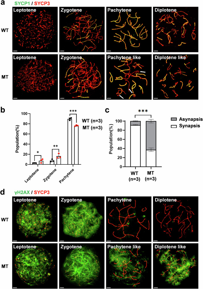
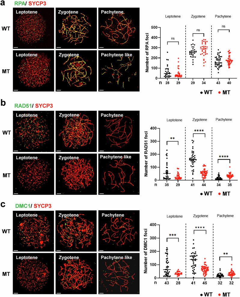
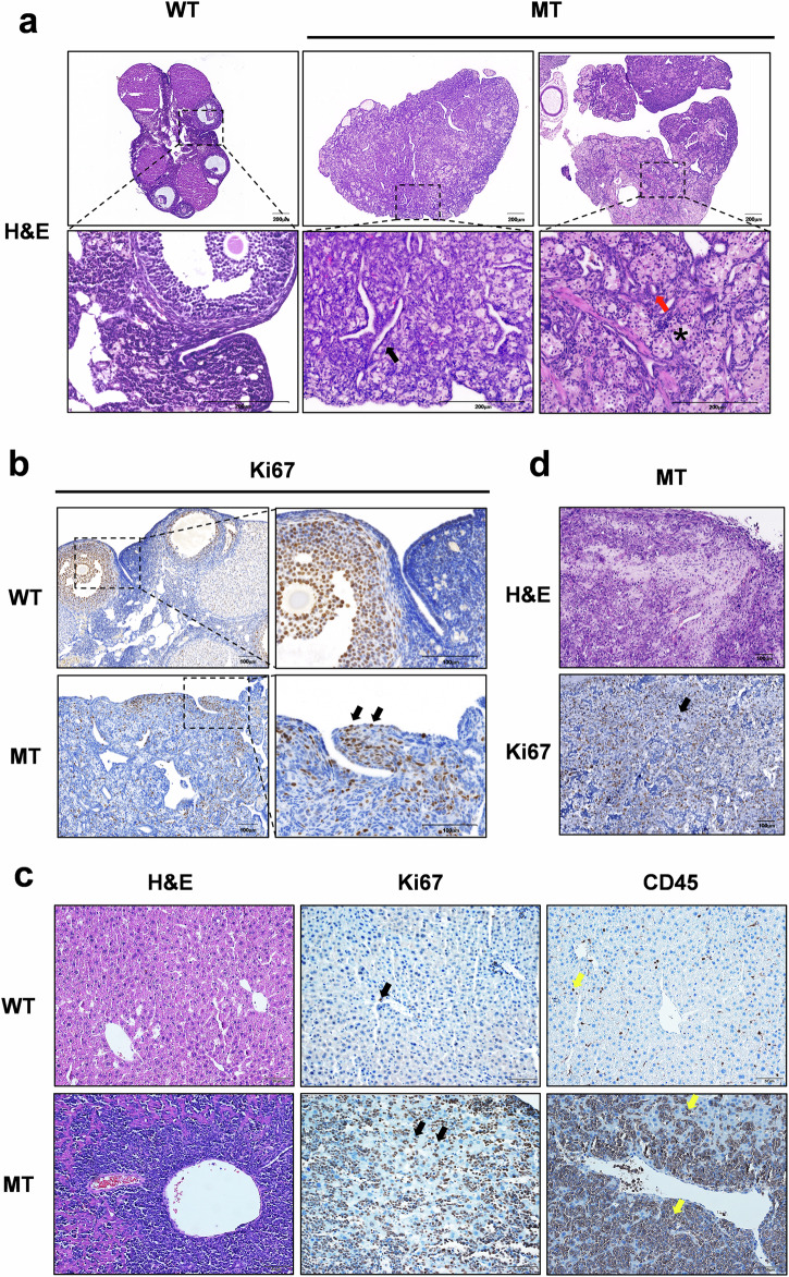
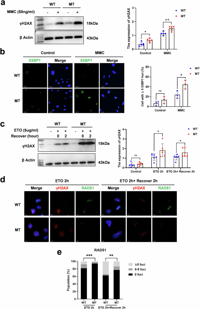
The DNA damage response plays a pivotal role in ovarian aging. Breast cancer susceptibility gene 2 (BRCA2), which participates in homologous recombination (HR), is a key regulator of natural menopause. Rare BRCA2 variants have been identified in patients with premature ovarian insufficiency (POI). However, the underlying molecular mechanisms are not well understood. Using a viable mouse model, Brca2c.68-1G>C/c.4384-4394del, carrying compound heterozygous variants mirroring the ones identified in a POI pedigree, we illustrated the essential role of BRCA2 in primordial follicle pool establishment. Germline deficiency of BRCA2 did not affect primordial germ cell (PGC) proliferation but impaired the recruitment of RAD51 and DMC1 to programmed DNA double-strand breaks (DSBs) during meiotic HR, causing postnatal oocyte depletion. Moreover, Brca2c.68-1G>C/c.4384-4394del mice presented increased tumor susceptibility. These findings confirmed the pathogenicity of BRCA2 biallelic variants in POI, revealing the dual impact on germ cell development and somatic cancer risk, underscoring the necessity of tumor surveillance in POI patients with BRCA2 mutations.


Conflict of interest statement

Competing interests: The authors declare no competing interests. Ethics statement: Ethical approval for the animal experiments was granted by the Institutional Review Board (IRB) of the Reproductive Medicine of Shandong University ([2014] IRB number: 52).

Figures

Fig. 1
Fig. 1. Accelerated oocyte consumption in the ovaries of Brca2c.68-1G>C/c.4384-4394del mice.
a Number of pups obtained by crossing Brca2c.68-1G>C/c.4384-4394del (MT) females with wild-type (WT) male mice for 3 months. Seven mice were analyzed in each group. b The size of the ovaries in 5-month-old (5 M) WT and MT females. c Hematoxylin and eosin staining of ovary sections from WT and MT mice at 21 days after birth (P21), 2-month-old (2 M), and 5 M. Black asterisks marked follicles in ovaries. Scale bars of P21 ovaries: 50 μm. Scale bars for 2 M and 5 M ovaries: 100 μm. d Immunofluorescence staining for STELLA or DDX4 and cleaved-PARP in gonad sections from embryonic day (E) 11.5, E18.5 and postnatal day (P) 0.5 WT and MT mice. Cell nuclei were counterstained with Hoechst (blue). White arrows showed the apoptotic germ cells in the gonads. Scale bars: 50 μm. e Quantitative analysis of STELLA/DDX4-positive oocyte numbers and the percentage of cleaved-PARP-positive oocytes per gonad. Data are presented as the mean ± SD and data points from individual groups are presented as dots. Ns, no statistically significant difference. ** P < 0.01. Student’s t-test.
Fig. 2
Fig. 2. Meiotic prophase I stage progression and synapsis in Brca2c.68-1G>C/c.4384-4394del oocytes.
a Immunofluorescence staining for SYCP1 and SYCP3 in leptotene, zygotene, pachytene, and diplotene oocytes from wild-type (WT) and Brca2c.68-1G>C/c.4384-4394del (MT) mice. The meiotic stages were determined based on SYCP3 signals. White arrows showed the chromosome with synaptic abnormality. Scale bars: 5 μm. b Percentage of oocytes at each stage of meiotic prophase. Three mice at E17.5 were analyzed in each group. Data are presented as mean ± SD. *, P < 0.05. ** P < 0.01. *** P < 0.001. Chi-square test, following Bonferroni correction. c Percentage of oocytes with or without synaptic abnormalities at the pachytene stage. Three mice at E17.5 were analyzed for each group. Data are presented as mean ± SD. ***, P < 0.001. Chi-square test. d Immunofluorescence staining for γ-H2AX and SYCP3 in leptotene, zygotene, pachytene, and diplotene oocytes from WT and MT mice. Scale bars: 5 μm.
Fig. 3
Fig. 3. Meiotic recombination in Brca2c.68-1G>C/c.4384-4394del oocytes.
Left: Oocytes from wild-type (WT) and Brca2c.68-1G>C/c.4384-4394del (MT) mice immunolabeled for RPA (a), RAD51 (b), and DMC1 (c) at leptotene, zygotene, and pachytene stages. Scale bars: 5 μm. Right: Quantification of the number of RPA (a), RAD51 (b), and DMC1 (c) that overlap with SYCP3 in each stage of WT and MT oocytes. The total number of analyzed oocytes is indicated. Data are presented as mean. Ns, no statistically significant difference. **, P < 0.01. ***, P < 0.001. ****, P < 0.0001. Student’s t test.
Fig. 4
Fig. 4. Increased tumor predisposition in Brca2c.68-1G>C/c.4384-4394del mice.
a Representative hematoxylin and eosin (H&E) staining of the ovary sections from wild-type (WT) female mice at 12 months and Brca2c.68-1G>C/c.4384-4394del (MT) female mice at 15 months, showing invagination of the germinal epithelium (black arrow), formation of isolated tubules (red arrow), and cell hyperplasia with luteinization (black asterisk) in the ovaries of MT mice. Dashed square indicated the area magnified. Scale bars: 200 μm. b Immunostaining with Ki67 (a proliferation marker) of the ovaries isolated from WT and MT mice illustrating moderate proliferative activity in the ovaries in MT females and a high density of Ki67-positive cells (black arrows) near the ovarian surface epithelium in MT mice. Black dashed lines marked magnified areas. Scale bars: 100 μm. c H&E and immunohistochemical staining of Ki67 and CD45 (a pan-leukocyte marker) in liver sections from WT and MT male mice at 7 months revealing high proliferative activity of Ki67 (black arrows) and a high density of CD45-positive cells (yellow arrows). Scale bars: 50 μm. d H&E and immunohistochemical staining against Ki67 in sarcoma in the leg of an MT male mouse at 14 months. Black arrow indicated the positive cell. Scale bars: 100 μm.
Fig. 5
Fig. 5. Adverse effect of Brca2 variants on DNA repair for DSBs in MEFs.
a Western blots of wild-type (WT) and Brca2c.68-1G>C/c.4384-4394del (MT) mouse embryonic fibroblasts (MEFs) for γH2AX and β-Actin (the loading control) following control and MMC (50 ng/ml) treatment for 24 h. b Percentage of the WT and MT MEFs with ≥5 53BP1 foci following control and MMC (50 ng/ml) treatment for 24 h. Three biological replicates were performed in each group and at least 150 MEFs were included per replicate. Scale bars: 20 μm. c Western blots of WT and MT MEFs for γH2AX and β-Actin following control, ETO (5 μg/ml) treatment for 2 h and then recovered for 2 h. d Representative images of RAD51 and γH2AX foci formation after ETO (5 μg/ml) treatment and 2-hour recovery in WT and MT MEFs. Scale bars: 7 μm. e Quantification of RAD51 foci seen in γH2AX -positive cells ( ≥ 5 foci). Data points from individual groups are presented as dots (a, b, c). Ns, no statistically significant difference. *, P < 0.05. **, P < 0.01. ***, P < 0.001. Student’s t test (a, b, c). Chi-square test (e).

Similar articles

References

    1. Christensen, K., Doblhammer, G., Rau, R. & Vaupel, J. W. Ageing populations: the challenges ahead. Lancet374, 1196–1208 (2009). - PMC - PubMed
    1. Inter LST Variations in reproductive events across life: a pooled analysis of data from 505 147 women across 10 countries. Hum. Reprod.34, 881–893 (2019). - PMC - PubMed
    1. Baird, D. T. et al. Fertility and ageing. Hum. Reprod. Update11, 261–276 (2005). - PubMed
    1. Ruth, K. S. et al. Genetic insights into biological mechanisms governing human ovarian ageing. Nature596, 393–397 (2021). - PMC - PubMed
    1. Lin, W., Titus, S., Moy, F., Ginsburg, E. S. & Oktay, K. Ovarian aging in women with BRCA germline mutations. J. Clin. Endocrinol. Metab.102, 3839–3847 (2017). - PMC - PubMed