. 2025 Jul 26;8(1):1111.
doi: 10.1038/s42003-025-08555-1.

Repression via DNA looping by the Gram-positive global transcriptional regulator ScoC from Geobacillus

Affiliations

Repression via DNA looping by the Gram-positive global transcriptional regulator ScoC from Geobacillus

Smadar Shulami et al. Commun Biol. .

Abstract

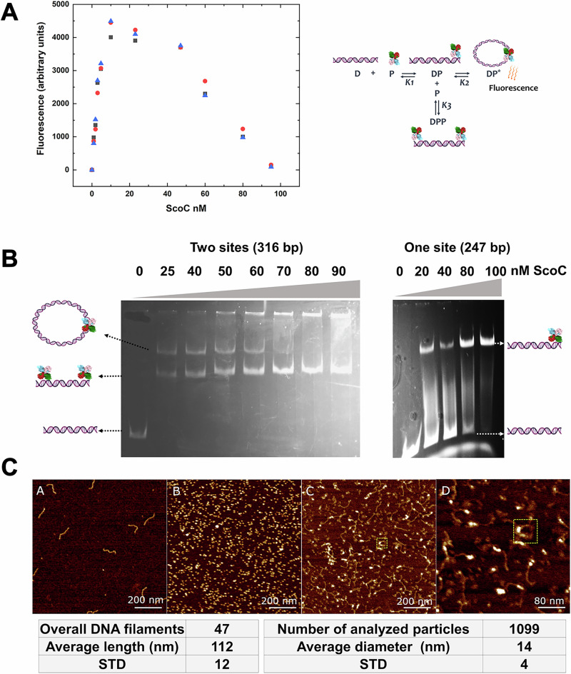
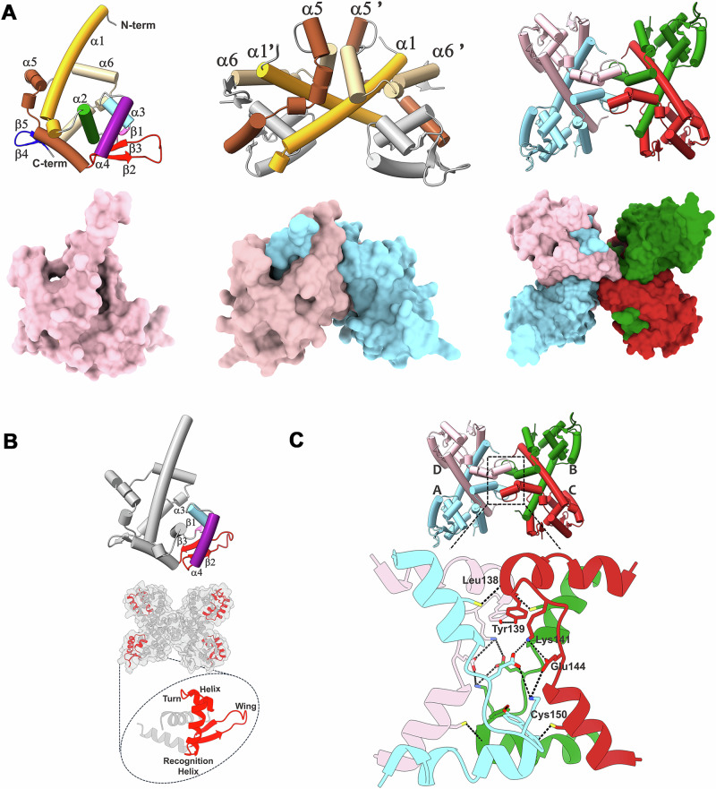
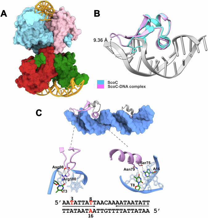
ScoC of the MarR family is a global regulator of transition phase pathways in Gram-positive bacteria, and in Bacillus subtilis it is estimated to regulate more than 500 genes. ScoC mediated activity is governed by its regulated expression, and by the interplay with other global transcriptional factors allowing for the finetuning of gene expression. Here we show, by transcriptional lacZ-fusions analysis, that ScoC from Geobacillus binds to two operator sites in the promoter region of the oligopeptide permease oppA, and that both binding sites are necessary for repression. Gel retardation assays, atomic force microscopy and fluorescence resonance energy transfer analyses demonstrate that ScoC can induce DNA looping. The crystal structures of ScoC and ScoC complexed with a 23-bp symmetric palindromic DNA from Geobacillus were determined at 3.15 Å and 3.50 Å resolution, respectively. The structures revealed a tetrameric X-shaped assembly composed of two dimers in which each dimeric unit comprises a winged helix-turn-helix DNA-binding motif. Our results expand the architecture of the MarR family regulators and suggest a mechanism by which ScoC interacts with other regulatory factors to modulate gene expression in the transition phase.


Conflict of interest statement

Competing interests: The authors declare no competing interests.

Figures

Fig. 1
Fig. 1. The oppA regulatory region contains two ScoC-binding sites, which are both required for repression.
A The ScoC-binding sequences are organized in two regions 208-bp apart. Each site is composed of two pseudo-palindromic sequences of 9-bases, and each of the 9-bases is also made of an internal pseudo-palindromic sequence. B Expression of oppA-lacZ-fusions demonstrating that two ScoC binding sites are required for repression. The left panel shows the truncated forms of the oppA regulatory regions. The mutated upstream ScoC-binding site I (ScoC I) contains specific substitutions of conserved nucleotides, indicated by underlined letters in the sequence: ATGATTAGTAACAATCATATAGT. The conserved nucleotides are presented in Supplementary Fig. 1. The right panel shows the LacZ activity of the different oppA-lacZ-fusions constructs. lacZ-fusions were constructed in B. subtilis SMY following chromosomal integration at the amyE locus. Cells were grown until mid-log in minimal medium (SMM) containing glucose, ammonium, and a mixture of 17 amino acids excluding Glu, His, and Tyr. β-Galactosidase (LacZ) activity was assayed and expressed in Miller units per cell turbidity at OD 600 nm. Numerical source data are in Supplementary Data 1.
Fig. 2
Fig. 2. ScoC induces DNA looping.
A FRET analysis demonstrating DNA looping mediated by ScoC. The fluorescence intensity was measured at 30 °C in the presence of different concentrations of ScoC with 10 nM of a 5’-Cy3 3’-Cy5 labeled 248-bp DNA element. The element was amplified from the oppA operator region and contained the two ScoC-binding sites at the ends. On the right is a cartoon demonstrating the equilibrium between the different intermediates, where D stands for DNA, and P stands for the tetrameric ScoC structure. At ScoC concentrations above 10 nM the equilibrium is shifted towards the DPP form, resulting in reduced fluorescence intensity. B Electrophoretic mobility shift assays demonstrating different DNA forms obtained upon binding of ScoC. Left panel: ScoC was incubated with a fluorescently labeled 316-bp DNA fragment containing two ScoC-binding sites at its ends. Two distinct retarded bands can be visualized. A slow migrating band representing DNA in the loop form (DP*) and a faster migrating band, representing the DNA in its linear form, in which two independent ScoC tetramers are bound at each end (DPP). In higher concentrations of ScoC only the fast-running band of the linear form appears. The DP form is not visible presumably since the equilibrium (K2) tends strongly towards the loop form (DP*). Right panel: a 247-bp DNA containing a single ScoC-binding site gives only a single retarded band shift representing a linear DNA form with one ScoC tetramer bound. Numerical source data are in Supplementary Data 2. C Visualizing ScoC-induced DNA looping by atomic force microscopy. AFM topography images of A 316 bp DNA fragment, B ScoC, C ScoC-DNA mixture showing different types of interactions and, D a zoom into a DNA loop induced by ScoC (yellow dashed square).
Fig. 3
Fig. 3. The three-dimensional structure of the ScoC.
A Ribbon representation of the monomer, dimer, and tetramer structures in which the topology of the MarR family is labeled. B The wing Helix turn Helix (wHTH) binding motif of ScoC. Each monomer of ScoC contains one motif unit shown in red. C The ScoC tetramerization contact region. The interactions forming the dimer-dimer interface include ten hydrogen bonds and four salt bridges involving residues: Tyr139, Lys141, Glu144, Cys150, and the backbone of Leu138.
Fig. 4
Fig. 4. ScoC is a tetramer in solution.
A Size-exclusion chromatogram of ScoC indicates that ScoC is a tetramer in solution with an apparent Mw of about 100,000 in solution. Protein standards used were Thyroglobulin (Mw, 669,000), Ferritin (Mw, 440,000), Aldolase (Mw, 158,000), Conalbumin (Mw, 75,000), and Ovalbumin (Mw, 44,000), all from GH Healthcare. Numerical source data are in Supplementary Data 3. B Scattering curve for ScoC. Log[I(q)] is plotted as a function of the momentum-transfer vector q. The q range is q = 0.007–0.037 Å−1. Experimental data are shown in black. The red curve corresponds to the simulated scattering curve for the envelope constructed using DAMMIN with χ2 = 0.95. The blue curve corresponds to the simulated scattering curve from the crystallographic tetrameric structure of ScoC calculated by CRYSOL with χ2 = 1.07. The graphs were generated using Origin 2019b (OriginLab Corporation, Northampton, MA, USA). Numerical source data are in Supplementary Data 4-6.
Fig. 5
Fig. 5. The crystal structure of the ScoC complexed with DNA.
A Each dimer binds a 23-bp palindrome dsDNA. The dimers are perpendicular to each other and consequently also the DNAs. B DNA binding to the ScoC winged HTH binding motif induces a 9.36 Å movement of the wing, allowing it to accommodate the minor groove. In blue is the free-winged HTH binding motif and in purple is the DNA-bound winged HTH binding motif. C Representation of the ScoC winged HTH binding motif occupying the minor and major grooves of the ScoC binding site. In the wing, occupying the minor groove, the conserved Arg100 is stabilized by Asp98 and interacts with the O2 atom of base T3. In the recognition helix, Asn79 interacts with the O4 atom of base T8 and Ser75 interacts with N6 of the complementary base, A16. The sequence of the ScoC binding site is shown at the bottom, and in red are the three bases that form hydrogen bonds with ScoC.

Similar articles

References

    1. Perego, M. & Hoch, J. A. Sequence analysis and regulation of the hpr locus, a regulatory gene for protease production and sporulation in Bacillus subtilis. J. Bacteriol.170, 2560–2567 (1988). - PMC - PubMed
    1. Adhya, S. Multipartite genetic control elements: communication by DNA loop. Annu. Rev. Genet.23, 227–250 (1989). - PubMed
    1. Amouyal, M. The remote control of transcription, DNA looping and DNA compaction. Biochimie73, 1261–1268 (1991). - PubMed
    1. Cournac, A. & Plumbridge, J. DNA looping in prokaryotes: experimental and theoretical approaches. J. Bacteriol.195, 1109–1119 (2013). - PMC - PubMed
    1. Saiz, L., Rubi, J. M. & Vilar, J. M. Inferring the in vivo looping properties of DNA. Proc. Natl. Acad. Sci. USA102, 17642–17645 (2005). - PMC - PubMed

MeSH terms

LinkOut - more resources