. 2025 Jul 27;15(1):27351.
doi: 10.1038/s41598-025-13237-3.

A designer polyQ fusion protein modulates NF-κB signaling by sequestering P65/RelA into aggregates

Affiliations

A designer polyQ fusion protein modulates NF-κB signaling by sequestering P65/RelA into aggregates

Xiang-Le Zhang et al. Sci Rep. .

Abstract

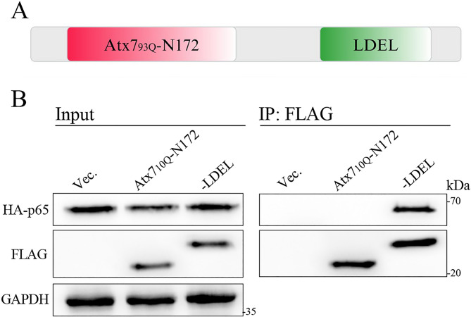
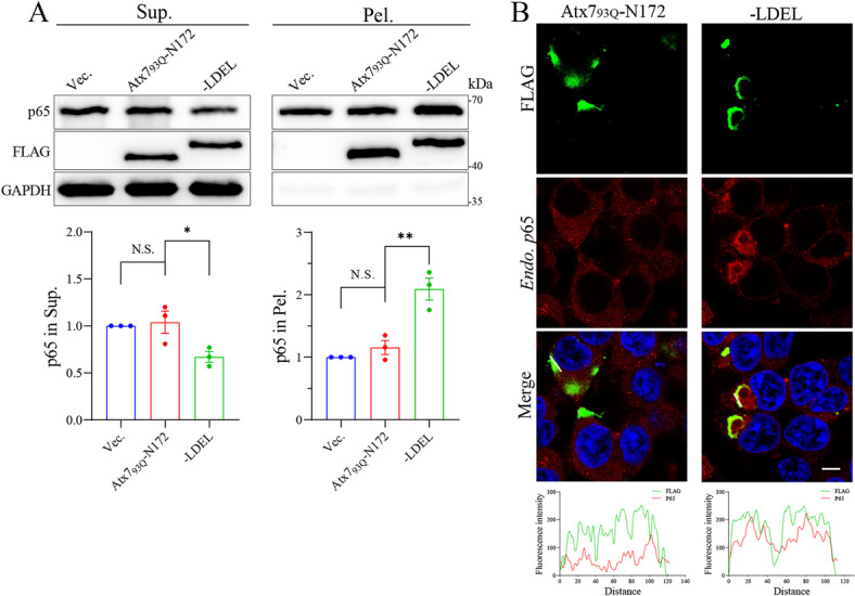
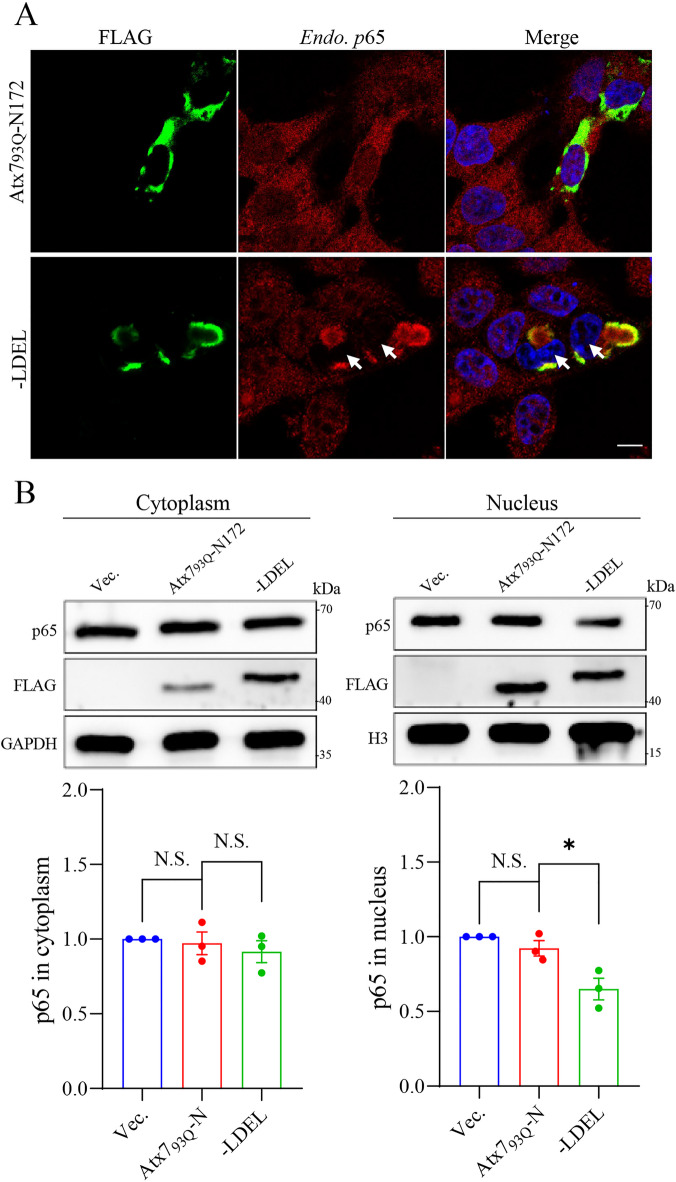
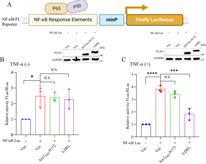
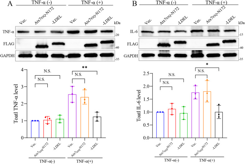
The transcription factor P65 is a subunit of the NF-κB complex that regulates expression of pro-inflammatory cytokines, thereby controlling the innate immune response. Excessive activation of NF-κB is commonly associated with various chronic inflammatory conditions. We previously established a polyglutamine (polyQ) fusion strategy to modulate biological processes by sequestering the targeted proteins. Here, we report a designer polyQ fusion (Atx793Q-N172-LDEL) that modulates the NF-κB signaling by sequestering P65 into aggregates. The fusion protein can interact with cellular P65 via its LDEL peptide sequence and specifically sequester it into aggregates or inclusions. This sequestration impedes the nuclear translocation process of P65, reduces its nuclear abundancy, and thereby attenuates the activity of NF-κB signaling and expression of the downstream genes, such as TNF-α and IL-6. This study provides therapeutic potential for treating inflammation and autoimmune disorders by targeting the P65 protein directly.

Keywords: Aggregation; LDEL peptide; NF-κB pathway.; P65 protein; PolyQ fusion; Sequestration.


Conflict of interest statement

Competing interests: The authors declare no competing interests.

Figures

Fig. 1
Fig. 1
Design of the polyQ-LDEL fusion and examination of its interaction with P65. (A) Schematic diagram of a designer polyQ fusion protein, Atx793Q-N172-LDEL. Atx793Q-N172, the N-terminal 172-residue fragment of Atx793Q. LDEL, a homologous peptide sequence, LDKALDELMDGDIIVFQK, from the UbL2 domain of USP7. (B) Examining the interaction between Atx710Q-N172-LDEL and P65 by co-IP assay. HEK 293T cells were co-transfected with FLAG-tagged Atx710Q-N172 or Atx710Q-N172-LDEL and HA-P65. At 48 h post-transfection, the cell lysates were subjected to co-IP assay using anti-FLAG agarose beads. Vec., vector; Atx710Q-N172, the N-terminal 172-residue fragment of Atx710Q; LDEL, Atx710Q-N172-LDEL, a polyQ fusion combined Atx710Q-N172 with LDEL.
Fig. 2
Fig. 2
The polyQ-LDEL fusion sequesters endogenous P65 into aggregates. (A) S/P fractionation for characterizing the distribution of endogenous P65 in supernatant and pellet affected by the polyQ-LDEL fusion. HEK 293T cells were transfected with FLAG-tagged Atx793Q-N172 or Atx793Q-N172-LDEL, followed by culturing for 48 h. The P65 levels in the supernatant and pellet fractions were then detected and analyzed. Sup., supernatant; Pel., pellet. Vec., vector; Atx793Q-N172, the N-terminal 172-residue fragment of Atx793Q; LDEL, Atx793Q-N172-LDEL, a polyQ fusion combined Atx793Q-N172 with LDEL. Data are shown as Mean ± SD (n = 3). *, p < 0.05; **, p < 0.01; N.S., no significance. (B) Immunofluorescence imaging for characterizing the co-localization of endogenous P65 with the inclusions formed by the polyQ-LDEL fusion. HEK 293T cells were transfected with FLAG-tagged Atx793Q-N172 or Atx793Q-N172-LDEL, and after cultured for 48 h, the cells were fixed and immunostained with anti-FLAG (green) and anti-P65 (red) antibodies. Nuclei were stained with Hoechst (blue). Scale bar = 10 μm. Bottom: co-localization analysis of the fluorescence signals for the distance represented by white lines.
Fig. 3
Fig. 3
The polyQ-LDEL fusion affects nuclear translocation of P65 in cells. (A) Immunofluorescence imaging for assessing the nuclear translocation of P65 affected by the polyQ-LDEL fusion. HEK 293T cells were transfected with FLAG-tagged Atx793Q-N172 or Atx793Q-N172-LDEL. After cultured for 48 h, the cells were treated with TNF-α (10 ng/mL) for 40 min. Subsequently, the cells were fixed and immunostained with anti-FLAG (green) and anti-P65 (red) antibodies. Nuclei were stained with Hoechst (blue). TNF-α, a cytokine for activating NF-κB signaling. Vec., vector; Atx793Q-N172, the N-terminal 172-residue fragment of Atx793Q; LDEL, Atx793Q-N172-LDEL. Scale bar = 10 μm. (B) Nuclear/cytoplasmic fractionation for assessing the nuclear translocation of P65 affected by the polyQ-LDEL fusion. HEK 293T cells were transfected with FLAG-tagged Atx793Q-N172 or Atx793Q-N172-LDEL. After cultured for 48 h, the cells were treated with TNF-α (10 ng/mL) for 40 min. Subsequently, the cells were collected and subjected to nuclear/cytoplasmic fractionation and detection of the P65 levels in the cytoplasm and nucleus. H3, histone 3. Data are presented as Mean ± SD (n = 3). *, p < 0.05; N.S., no significance.
Fig. 4
Fig. 4
The polyQ-LDEL fusion modulates the NF-κB signaling activity. (A) Schematic diagram of the NF-κB-FL reporter. The NF-κB response element harbors a DNA sequence of tandem GGGAATTTCC, which drives the expression of the Firefly luciferase gene through the mini-promoter on the plasmid. minP, mini-promoter. (B, C) Effects of the polyQ-LDEL fusion on the luciferase activities. The pGL3-NF-κB-FL plasmid as well as the pGL3 vector harboring a Renilla luciferase gene was co-transfected with FLAG-tagged Atx793Q-N172 or Atx793Q-N172-LDEL into HEK 293T cells. After cultured for 48 h, the cells were treated without (B) or with (C) TNF-α (10 ng/mL) for 40 min, and then the cell lysates were subjected to luciferase activity assay (FLuc/RLuc). The empty vector was set as a control. Insert: immunoblots showing the expression levels of Atx793Q-N172 and its LDEL-fused form. Vec., vector; Atx793Q-N172, the N-terminal 172-residue fragment of Atx793Q; LDEL, Atx793Q-N172-LDEL. Data are presented as Mean ± SD (n = 3). *, p < 0.05; ***, p < 0.001; ****, p < 0.001; N.S., no significance.
Fig. 5
Fig. 5
Effects of the polyQ-LDEL fusion on the downstream gene expression. TNF-α expression. (B) IL-6 expression. HEK 293T cells were transfected with FLAG-tagged Atx793Q-N172 or Atx793Q-N172-LDEL. After cultured for 30 h, the cells were treated with TNFα (10 ng/mL) for 40 min. The cell lysates were then subjected to immunoblotting with an anti-TNF-α or anti-IL6 antibody to assess the protein level of TNF-α (A) or IL-6 (B). Vec., vector; Atx793Q-N172, the N-terminal 172-residue fragment of Atx793Q; LDEL, Atx793Q-N172-LDEL. Data are presented as Mean ± SD (n = 3). *, p < 0.05; **, p < 0.01; N.S., no significance.

Similar articles

References

    1. Housmans, J. A. J., Wu, G., Schymkowitz, J. & Rousseau, F. A guide to studying protein aggregation. FEBS J.290, 554–583. 10.1111/febs.16312 (2023). - PubMed
    1. Chiti, F. & Dobson, C. M. Protein misfolding amyloid formation, and human disease: a summary of progress over the last decade. Annu. Rev. Biochem.86, 27–68. 10.1146/annurev-biochem-061516-045115 (2017). - PubMed
    1. Lieberman, A. P., Shakkottai, V. G. & Albin, R. L. Polyglutamine repeats in neurodegenerative diseases. Annu. Rev. Pathol.14, 1–27. 10.1146/annurev-pathmechdis-012418-012857 (2019). - PMC - PubMed
    1. Arrasate, M., Mitra, S., Schweitzer, E. S., Segal, M. R. & Finkbeiner, S. Inclusion body formation reduces levels of mutant Huntingtin and the risk of neuronal death. Nature431, 805–810. 10.1038/nature02998 (2004). - PubMed
    1. Mahul-Mellier, A. L. et al. The process of lewy body formation, rather than simply alpha-synuclein fibrillization, is one of the major drivers of neurodegeneration. Proc. Natl. Acad. Sci. U S A. 117, 4971–4982. 10.1073/pnas.1913904117 (2020). - PMC - PubMed

MeSH terms

LinkOut - more resources