. 2025 Dec;17(1):2537753.
doi: 10.1080/19490976.2025.2537753. Epub 2025 Jul 28.

Dietary cholesterol impairs cognition via gut microbiota-derived deoxycholic acid in obese mice

Affiliations

Dietary cholesterol impairs cognition via gut microbiota-derived deoxycholic acid in obese mice

Yan Liu et al. Gut Microbes. 2025 Dec.

Abstract

Dietary cholesterol is often found in a high-fat diet (HFD) and excessive intake is harmful to cognitive function. The gut microbiome constitutes an environmental factor influenced by diet, which regulates cognitive function via the gut-brain axis. The present study explored the role of dietary cholesterol in HFD-induced cognitive impairment and the participation of the gut microbiota and metabolites. Here, we found that dietary cholesterol promoted cognitive impairment in HFD-fed mice, which was associated with an increase in gut microbiota containing 7α-dehydroxylase, including Lachnospiraceae bacterium, Dorea sp. Clostridium sp. and elevated levels of deoxycholic acid (DCA) in the hippocampus. Upon dietary cholesterol intake, the activity of gut microbiota in mice to produce DCA is increased. Fecal microbiota transplantation confirmed that the cognitive impairment-promoting process was driven by gut microbiota. Reducing circulating bile acid levels with cholestyramine improved cognitive decline in mice, whereas hippocampal administration of DCA worsened cognitive function. Pharmacological inhibition of hippocampal apical sodium bile acid transporter reduces neuronal DCA accumulation and improves neuronal apoptosis as well as cognitive impairments in mice. Overall, this study revealed that dietary cholesterol promotes HFD-induced cognitive impairment by inducing the production of DCA through gut microbiota metabolism.

Keywords: Dietary cholesterol; bile acid; cognitive impairment; deoxycholic acid; gut microbiota.


Conflict of interest statement

No potential conflict of interest was reported by the author(s).

Figures

Figure 1.
Figure 1.
Dietary cholesterol promotes cognitive impairment in HFD-fed mice. (A) experimental framework. Mice were fed with a control, HFD, and HFD with various doses of cholesterol (0.2%, 0.5%, and 1%) for 6 months. Then, Y-maze and nor tests were performed on each mouse. (B) body weight change (n = 10). (C) serum lipids profile (n = 6). (D) percentage of alteration and total arm entries in the Y-maze test (n = 10). (E) discrimination rate and total exploration time in the nor test (n = 10). Statistical analysis was performed using a one-way ANOVA followed by Dunnett post hoc test. * p < 0.05, ** p < 0.01, *** p < 0.001.
Figure 2.
Figure 2.
Apoptosis and bile acid transportation pathways were activated in dietary cholesterol-fed mice. (A) Principal component analysis (PCA) of mouse hippocampal transcriptome profile based on princomp function. (B) Volcano map based on DEGs (padj < 0.05 and Fold change ≥ 1.5 or Fold change ≤ 0.67). (C) GSEA analysis of apoptosis signaling pathway enriched in KEGG analysis (n = 3). (D) GSEA analysis of positive regulation of neuron apoptotic process in GO analysis (n = 3). (E) GSEA analysis of bile acid and bile salt transport in GO analysis (n = 3). (F) expression levels of genes associated with apoptosis based on qPCR tests (n = 6). (G) expression levels of genes associated with bile acid and bile salt transport (Slc10a2) (n = 6). (H) expression of proteins associated with apoptosis (cleaved caspase 3) based on Western blot (n = 3). (I) expression of proteins related to bile acid and bile salt transport (ASBT) based on Western blot (n = 3). (J) Representative micrographs of hippocampal tissue stained with H&E (magnification 100×). (K) the assessment of apoptotic cell numbers within the hippocampal region (n = 6). Statistical analysis was performed using a one-way ANOVA followed by Dunnett post hoc test. * p < 0.05, ** p < 0.01, *** p < 0.001.
Figure 3.
Figure 3.
Dietary cholesterol increased fecal microbiota containing 7ɑ-dehydroxylase and hippocampal DCA in HFD-fed mice. (A) PLS-DA analysis of metabolic profile in the HFD and HFD + 0.2%Chol groups (n = 5). (B) volcano plots illustrate variations in metabolite levels in murine serum (n = 5). (C) hippocampal levels of unconjugated and taurine-conjugated BAs (n = 5). (D) PCoA of fecal microbiota profile in the HFD and HFD + 0.2%Chol groups at the species level (n = 6). (E) ɑ-diversity of fecal microbiota in the HFD and HFD + 0.2%Chol groups at the species level (n = 6). (F) relative abundance of lachnospiraceae bacterium, Dorea sp., Clostridium sp., and Eubacterium sp. in murine feces (n = 6). (G) reads number of cbh in fecal microbiota (n = 6). (H) fecal BSH activity in mice (n = 6). (I) reads number of baiN in fecal microbiota (n = 6). (J) in vitro assessment of DCA production by fecal bacteria (n = 6). For the differences of microbiota, the Wilcoxon rank-sum test (two-tailed test) was employed. For the other data, an unpaired t-test or one-way ANOVA followed by Dunnett post hoc test was used. * p < 0.05, ** p < 0.01, *** p < 0.001.
Figure 4.
Figure 4.
Pro-cognitive impairment effects of dietary cholesterol can be transferred by gut microbiota. (A) experimental framework. (B) body weight change (n = 10). (C) percentage of alteration and total arm entries in the Y-maze test (n = 10). (D) discrimination rate and total exploration time in the nor test (n = 10). (E) Representative micrographs of hippocampal tissue stained with H&E (magnification 100×). (F) the assessment of apoptotic cell numbers within the hippocampal region (n = 6). (G) protein level of C-Caspase 3 (n = 3). (H) protein expression of ASBT (n = 3). Statistical analysis was performed using one-way ANOVA followed by Dunnett post hoc test. * p < 0.05, ** p < 0.01, *** p < 0.001.
Figure 5.
Figure 5.
Deoxycholic acid-induced cognitive impairment in HFD-fed mice. (A) experimental framework. (B) percentage of alteration and total arm entries in the Y-maze test (n = 10). (C) discrimination rate and total exploration time in the nor test (n = 10). (D) Representative micrographs of hippocampal tissue stained with H&E (magnification 100×) and the assessment of apoptotic cell numbers within the hippocampal region (n = 6). (E) experimental framework. (F) percentage of alteration and total arm entries in the Y-maze test (n = 10). (G) discrimination rate and total exploration time in the nor test (n = 10). (H) Representative micrographs of hippocampal tissue stained with H&E (magnification 100×) and the assessment of apoptotic cell numbers within the hippocampal region (n = 6). Statistical analysis was performed using an unpaired t-test. * p < 0.05, ** p < 0.01, *** p < 0.001.
Figure 6.
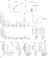
Figure 6.
ASBT regulates DCA-induced neuronal apoptosis. (A) cell viability after 25–75 μM DCA treatments in SH-SY5Y for 24 h (n = 6). (B) TUNEL density after 25–75 μM DCA treatments in SH-SY5Y for 24 h (n = 6). (C) JC-1 monomer/aggregation after 25–75 μM DCA treatments in SH-SY5Y for 12 h (n = 6). (D) dynamics of caspase 3 activity after 25–75 μM DCA treatments in SH-SY5Y for 24 h (n = 6). (E) TUNEL density after DEX (1–10 μM) and DCA (25 μM) co-treatments in SH-SY5Y for 96 h (n = 6). (F) TUNEL density after GSK (5–25 μM) and DCA (75 μM) co-treatments in SH-SY5Y for 24 h (n = 6). (G) experimental design. (H) effects of GSK on hippocampal DCA level (n = 6). (I) percentage of alteration and total arm entries in the Y-maze test (n = 10). (J) discrimination rate and total exploration time in the novel object recognition test (n = 10). (K) Representative images of hippocampus H&E staining (magnification 100×) and the quantification of apoptotic cells in the hippocampus (n = 6). (L) effects of DCA on the number of apoptotic cells in different regions of the mouse hippocampus (n = 6). (M) protein expression of cleaved-caspase-3 (n = 3). (N) schematic diagram of the mechanism of dietary cholesterol promotes HFD-induced cognitive impairment. In (a-c) and (e-f), statistical analysis was performed using one-way ANOVA followed by Dunnett post hoc test. In (D) and (H-L), statistical analysis was performed using two-way ANOVA followed by Tukey post hoc test. * p < 0.05, ** p < 0.01, *** p < 0.001. DEX, dexamethasone; GSK, GSK2330672.

References

    1. Liu Y, Xiao H, Wang Z, Pan Q, Zha X, Lu B. Interactions between dietary cholesterol and intestinal flora and their effects on host health. Crit Rev Food Sci Nutr. 2025;65(3):494–26. doi: 10.1080/10408398.2023.2276883. - DOI - PubMed
    1. Zhang T, Xie L, Guo Y, Wang Z, Guo X, Lu R, Jin Q, Chang M, Wang X. 4,4-dimethylsterols reduces fat accumulation via inhibiting fatty acid amide hydrolase in vitro and in vivo. Research. 2024;7:0377. doi: 10.34133/research.0377. - DOI - PMC - PubMed
    1. Zhu H, Chen Z-Y.. Do we No longer need to worry about dietary cholesterol? J Agr Food Chem. 2017;65(46):9931–9933. doi: 10.1021/acs.jafc.7b04829. - DOI - PubMed
    1. Zhong VW, Van Horn L, Cornelis MC, Wilkins JT, Ning, H, Carnethon, MR, Greenland P, Mentz RJ, Tucker KL, Zhao L, et al. Associations of dietary cholesterol or egg consumption with incident cardiovascular disease and mortality. J Sport Hist American Medical Association. 2019;321(11):1081–1095. doi: 10.1001/jama.2019.1572. - DOI - PMC - PubMed
    1. Noureddin M, Zelber‐Sagi S, Wilkens LR, Porcel J, Boushey CJ, Le Marchand L, Rosen HR, Setiawan VW. Diet associations with nonalcoholic fatty liver disease in an ethnically diverse population: the multiethnic cohort. Hepatology. 2020;71(6):1940–1952. doi: 10.1002/hep.30967. - DOI - PMC - PubMed

MeSH terms

LinkOut - more resources