Review
. 2025 Jun 16:12:rbaf059.
doi: 10.1093/rb/rbaf059. eCollection 2025.

Engineering of tissue in microphysiological systems demonstrated by modelling skeletal muscle

Affiliations
Review

Engineering of tissue in microphysiological systems demonstrated by modelling skeletal muscle

Yuan Gao et al. Regen Biomater. .

Abstract

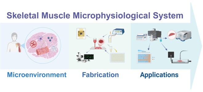
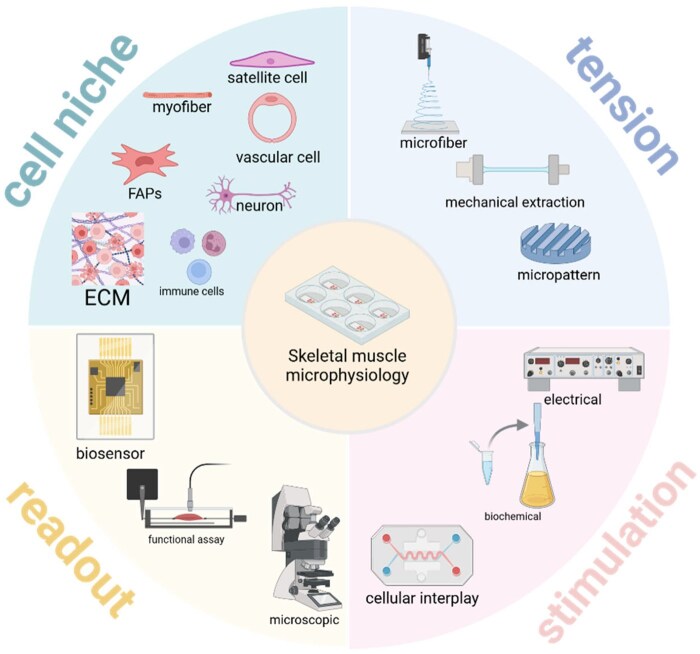
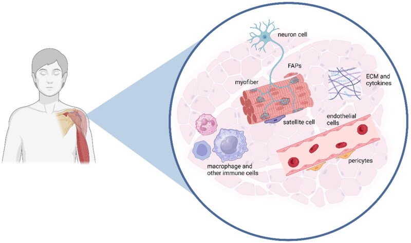
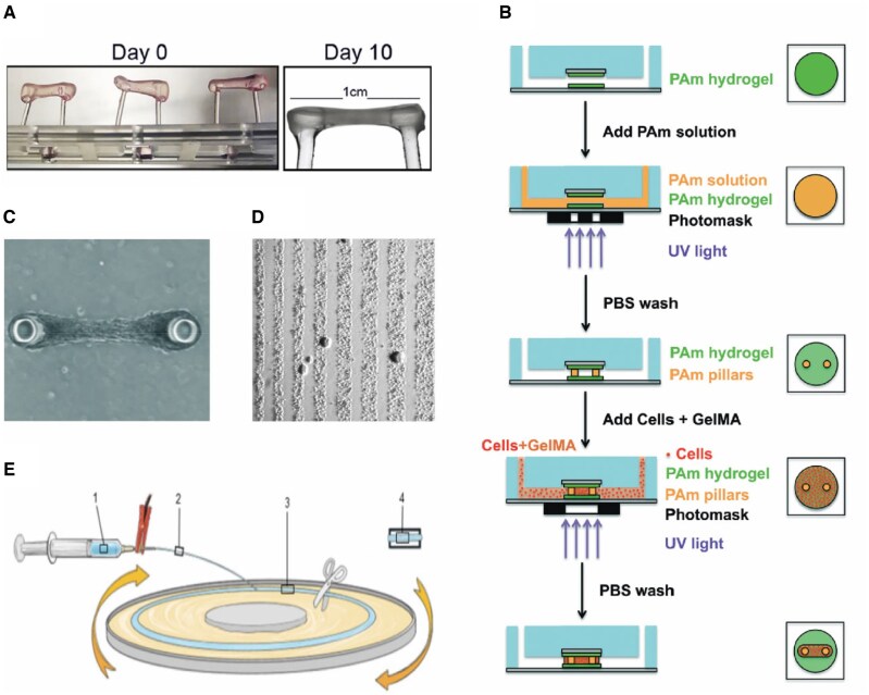
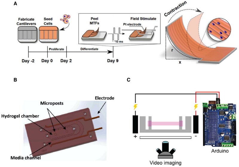
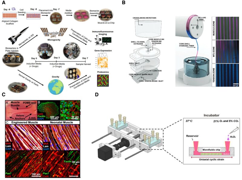
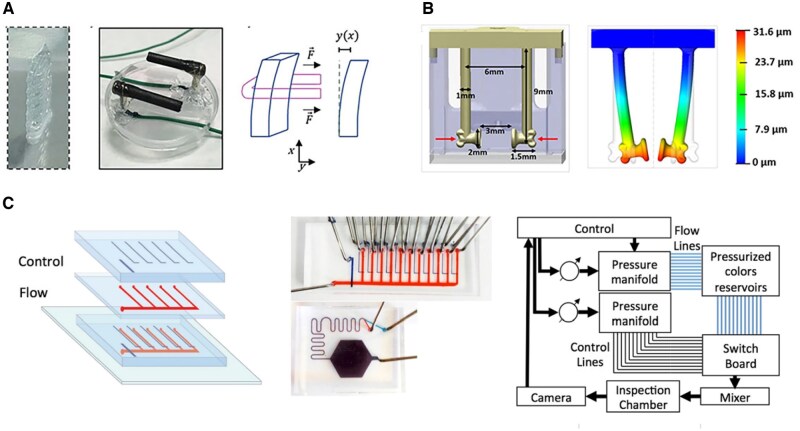
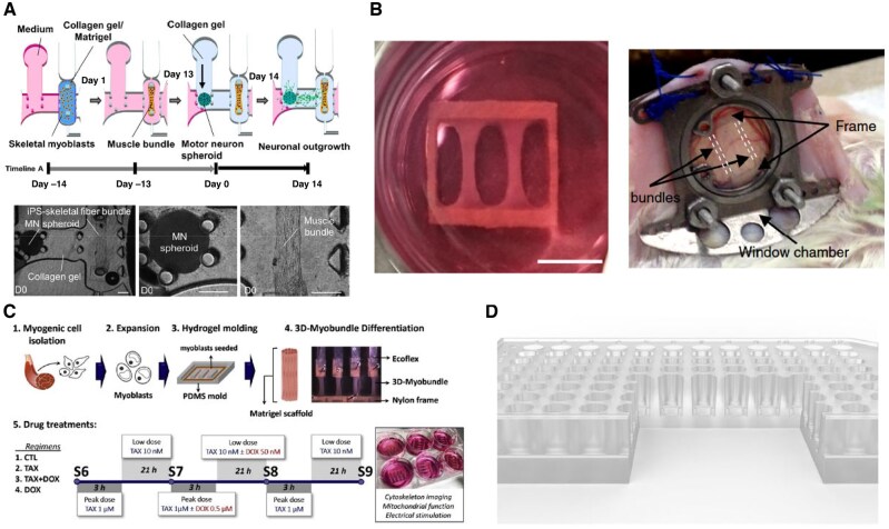
Research on myogenesis and myogenic pathologies has garnered significant attention in recent years. However, traditional in vitro modeling approaches have struggled to fully replicate the complex functions of skeletal muscle. This limitation is primarily due to the insufficient reconstruction of the muscle tissue microenvironment and the role of physical cues in regulating muscle cell activity. Recent studies have highlighted the importance of the microenvironment, which includes cells, extracellular matrix (ECM) and cytokines, in influencing myogenesis, regeneration and inflammation. This review focuses on advances in skeletal muscle construction toward a complete microphysiological system, such as organoids and muscle-on-a-chip technology, as well as innovative interventions like bioprinting and electrical stimulation. These advancements have enabled researchers to restore functional skeletal muscle tissue, bringing us closer to achieving a fully functional microphysiological system. Compared to traditional models, these systems allow for the collection of more comprehensive data, providing insights across multiple scales. Researchers can now study skeletal muscle and disease models in vitro with increased precision, enabling more advanced research into the physiological and biochemical cues affecting skeletal muscle activity. With these advancements, new applications are emerging, including drug screening, disease modeling and the development of artificial tissues. Progression in this field holds great promise for advancing our understanding of skeletal muscle function and its associated pathologies, offering potential therapeutic solutions for a variety of muscle-related diseases.

Keywords: bioengineering; in vitro modeling; microenvironment; microphysiological system; skeletal muscle.


Figures

None
Graphical abstract
Figure 1.
Figure 1.
Engineering of skeletal muscle tissue in microphysiological system. Created in BioRender. gao, y. (2025).
Figure 2.
Figure 2.
The microenvironment of skeletal muscle. Created in BioRender. gao, y. (2025). The microenvironment of skeletal muscle includes muscle progenitor cells like satellite cells, extracellular matrix, blood vessels, immune cells and other components. A stable cellular microenvironment ensures normal myocyte function, maintains satellite cells quiescent and is jointly regulated by internal and external factors.
Figure 3.
Figure 3.
Planar culture and organoid models of skeletal muscle. (A) Myoblast colonies differentiated from hPSCs in a planar culture system over different time points during the process, the number in each section shows the time order [46]. (B) Schematic of co-culture system for human motor neurons and skeletal muscle cells in a Transwell model for neuromuscular junction formation. hSpS: human spinal cord cells, hSkM: human skeletal muscle cells [47]. (C) A protocol scheme for inducing hPSC differentiation into myogenic progenitor cells using small molecules, divided into three distinct phases: paraxial mesoderm induction, myogenic induction and myogenic growth [48].
Figure 4.
Figure 4.
Different attempts to introduce tension into muscle microphysiological systems. (A) Schematic diagram of applying tension to myoblasts encapsulated in a hydrogel to induce myofascial generation [55]. (B) Process of fabrication of a muscle-on-chip device with two pillars set up to provide fulcrums for myogenic cells, which utilize various photo-curable materials for model fabrication. PAm: acrylamide (Am) solution containing photoinitiator, GelMA: gelatin methacrylate [73]. (C) Microscopic images of the muscle-on-a-chip device after culture maturation, showing a bundle-like structure [73]. (D) Directing myofibrillar generation by sculpting surface patterns to provide surface tension to myocytes [84]. (E) Schematic of microfiber production for muscle cell alignment, involving cell and hydrogel mixture loading into a syringe, extrusion under applied voltage, fiber formation on a rotating platform and final collection [80].
Figure 5.
Figure 5.
Introduction of electrical stimulation and activation of muscle to the skeletal muscle microphysiological system. (A) Cultured muscle tissue was placed into an electric field to induce muscle contraction [82]. (B) Electrodes were introduced into the microfluidic chip to create organoids to simulate muscle contraction [77]. (C) Electrodes were inserted into the cultured complete muscle bundles, and the cyclic electrical stimulation inputs were fed through the artificial circuitry to emulate the muscle physiological environment [70].
Figure 6.
Figure 6.
Introduction of other physiological and biochemical factors into skeletal muscle microphysiological systems. (A) Fabrication of organoids by microfiber-guided muscle generation and sending them to microgravity environments for comparative studies in culture, comparing functional and pharmacological validation [99]. (B) Obtaining musculotendinous junction tissues by hybrid 3D printing [56]. (C) Introduction of endothelial cells for the cultivation of vascularized artificial muscle bundles [107]. (D) Introduction of mechanically and chemically damaged microphysiological systems [74].
Figure 7.
Figure 7.
Detection of various types of signals in the microphysiological system. (A) Electrical stimulation triggers muscle contraction, pulling on the basal column deformation and converting tension by pre-measured modulus of elasticity [85]. (B) Quantitative measurements of the basal column deformation induced by the muscle pulling [126]. (C) Introducing a variety of sensors into the microfluidic system and participating in its modulation [127].
Figure 8.
Figure 8.
Different application scenarios of the artificial muscle microphysiology system. (A) Establishment of NMJ organ chips for simulating diseases such as ALS and drug screening [76]. (B) Cultivated artificial muscle bundles can also be reimplanted in vivo to validate their physiological function processes [57]. (C) Microchip models for validating the toxicity of chemotherapeutic drugs on muscle tissue [88]. (D) High-throughput muscle microchip models that can be used to carry out experiments such as drug screening in a more efficient manner [69].

Similar articles

  • Short-Term Memory Impairment.
    Cascella M, Al Khalili Y. Cascella M, et al. 2024 Jun 8. In: StatPearls [Internet]. Treasure Island (FL): StatPearls Publishing; 2025 Jan–. 2024 Jun 8. In: StatPearls [Internet]. Treasure Island (FL): StatPearls Publishing; 2025 Jan–. PMID: 31424720 Free Books & Documents.
  • Management of urinary stones by experts in stone disease (ESD 2025).
    Papatsoris A, Geavlete B, Radavoi GD, Alameedee M, Almusafer M, Ather MH, Budia A, Cumpanas AA, Kiremi MC, Dellis A, Elhowairis M, Galán-Llopis JA, Geavlete P, Guimerà Garcia J, Isern B, Jinga V, Lopez JM, Mainez JA, Mitsogiannis I, Mora Christian J, Moussa M, Multescu R, Oguz Acar Y, Petkova K, Piñero A, Popov E, Ramos Cebrian M, Rascu S, Siener R, Sountoulides P, Stamatelou K, Syed J, Trinchieri A. Papatsoris A, et al. Arch Ital Urol Androl. 2025 Jun 30;97(2):14085. doi: 10.4081/aiua.2025.14085. Epub 2025 Jun 30. Arch Ital Urol Androl. 2025. PMID: 40583613 Review.
  • The Lived Experience of Autistic Adults in Employment: A Systematic Search and Synthesis.
    Thorpe D, McKinlay M, Richards J, Sang K, Stewart ME. Thorpe D, et al. Autism Adulthood. 2024 Dec 2;6(4):495-509. doi: 10.1089/aut.2022.0114. eCollection 2024 Dec. Autism Adulthood. 2024. PMID: 40018061 Review.
  • The Black Book of Psychotropic Dosing and Monitoring.
    DeBattista C, Schatzberg AF. DeBattista C, et al. Psychopharmacol Bull. 2024 Jul 8;54(3):8-59. Psychopharmacol Bull. 2024. PMID: 38993656 Free PMC article. Review.
  • Innovative organ-on-a-chip platforms for exploring tumorigenesis and therapy in head and neck cancer.
    Lin C, Zhang Z, Yang F, Gu S, Zuo J, Wu Y, Zhang J, Zhou T, Zhang Y, Chen Z, Gu Z, Shen Z. Lin C, et al. J Transl Med. 2025 Jul 16;23(1):798. doi: 10.1186/s12967-025-06824-5. J Transl Med. 2025. PMID: 40671128 Free PMC article. Review.

References

    1. Mukund K, Subramaniam S. Skeletal muscle: a review of molecular structure and function, in health and disease. WIREs Syst Biol Med 2020;12:e1462. - PMC - PubMed
    1. Rodríguez C, Timóteo-Ferreira F, Minchiotti G, Brunelli S, Guardiola O. Cellular interactions and microenvironment dynamics in skeletal muscle regeneration and disease. Front Cell Dev Biol 2024;12:1385399. - PMC - PubMed
    1. Dumont NA, Wang YX, Rudnicki MA. Intrinsic and extrinsic mechanisms regulating satellite cell function. Development 2015;142:1572–81. 10.1242/dev.114223. - DOI - PMC - PubMed
    1. Sousa-Victor P, García-Prat L, Muñoz-Cánoves P. Control of satellite cell function in muscle regeneration and its disruption in ageing. Nat Rev Mol Cell Biol 2022;23:204–26. - PubMed
    1. Guo Q, Luo Q, Song G. Control of muscle satellite cell function by specific exercise‐induced cytokines and their applications in muscle maintenance. J Cachexia Sarcopenia Muscle 2024;15:466–76. - PMC - PubMed

LinkOut - more resources