. 2025 Jul 12:53:100701.
doi: 10.1016/j.jbo.2025.100701. eCollection 2025 Aug.

Yin Yang 1 protein-activated N-acetyltransferase 10 drives cell malignant progression of osteosarcoma through ac4C acetylation of integrin β3

Affiliations

Yin Yang 1 protein-activated N-acetyltransferase 10 drives cell malignant progression of osteosarcoma through ac4C acetylation of integrin β3

Fan Yang et al. J Bone Oncol. .

Abstract

Background: N-acetyltransferase 10 (NAT10) acts as the "writer" of N4-acetylcytidine (ac4C) modification in tumor progression, including osteosarcoma (OS). Its molecular mechanism in OS remains not fully clear. This study endeavored to disclose the upstream and downstream mechanism of NAT10 related to Yin Yang 1 protein (YY1) and integrin β3 (ITGB3) in OS.

Methods: Gene mRNA and protein levels were assayed via real-time quantitative PCR and Western blotting. Cell counting kit-8, EdU assay, flow cytometry/TUNEL staining assay, transwell assay, and scratch assay were conducted to assess cell viability, proliferation, apoptosis, invasion, and migration. Interaction analysis was completed through ac4C RNA immunoprecipitation (ac4c RIP), RIP, chromatin IP and dual-luciferase reporter assay. In vivo assay was carried out using xenograft models in mice.

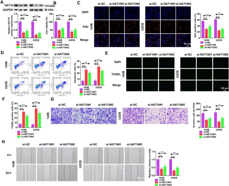
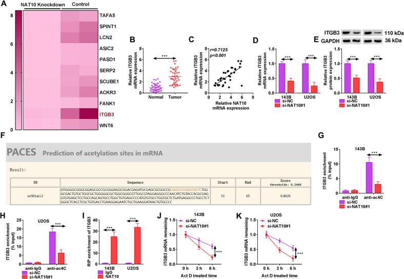
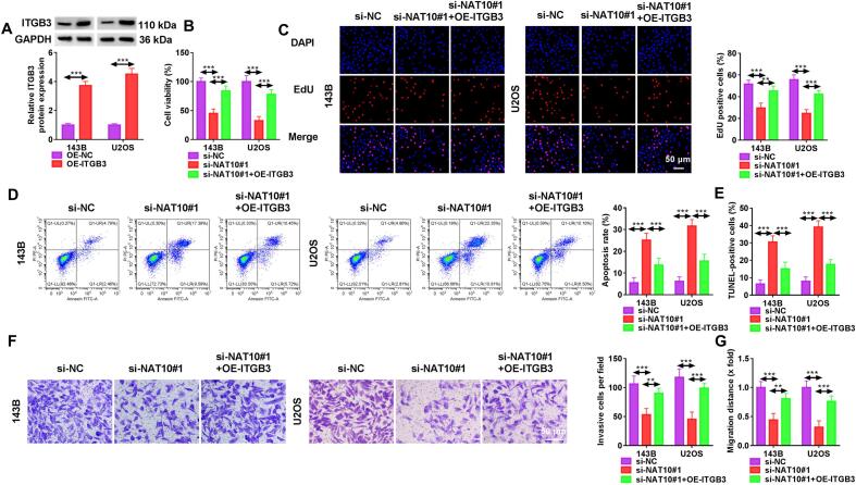
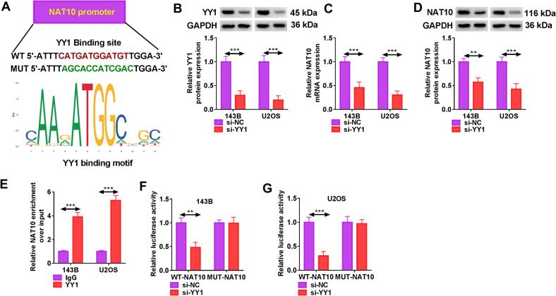
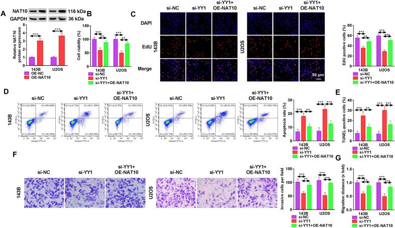
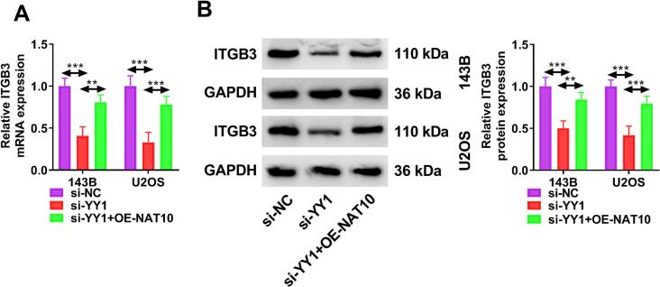
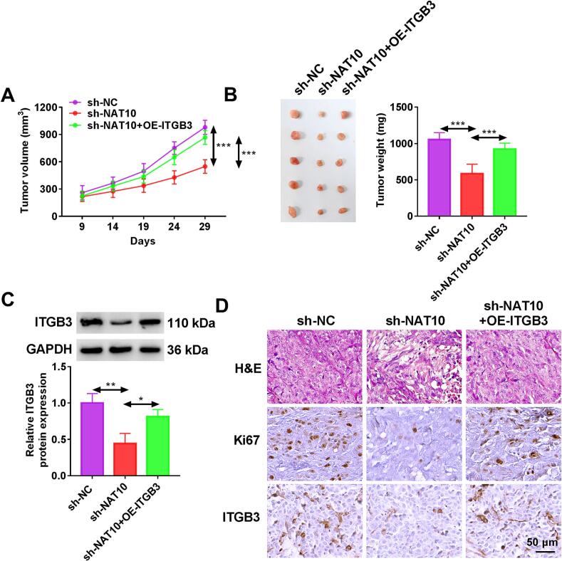
Results: OS tissues and cells showed the high expression of NAT10. Cell proliferation, invasion, and migration were suppressed but apoptosis was enhanced in NAT10-silenced OS cells. GSE237541 dataset has predicted the inhibition of ITGB3 after NAT10 knockdown, and PACES website predicted ac4C site in ITGB3. Furthermore, it was found that NAT10 could up-regulate ITGB3 expression by mediating ac4C acetylation. ITGB3 overexpression recused OS cell progression inhibition caused by NAT10 knockdown. Jaspar predicted the binding between YY1 and NAT10 promoter. YY1 could activate the transcriptional regulation of NAT10 to increase NAT10 expression, and YY1 depletion blocked cell malignant behaviors via reducing NAT10 expression. More importantly, YY1 interacted with NAT10 to up-regulate ITGB3 expression. In vivo, NAT10/ITGB3 axis also promoted OS tumor growth in mice.

Conclusion: YY1 was firstly affirmed to regulate transcription of NAT10, and NAT10 was firstly indicated to mediate ac4C modification of ITGB3. YY1-activated NAT10 could affect ITGB3 and then modulated the malignant development of OS.

Keywords: Integrin β3; N-acetyltransferase 10; Osteosarcoma; Yin Yang 1 protein; ac4C acetylation.


Conflict of interest statement

The authors declare that they have no known competing financial interests or personal relationships that could have appeared to influence the work reported in this paper.

Figures

None
Graphical abstract
Fig. 1
Fig. 1
OS tissues and cells expressed NAT10 with a high level. (A-B) NAT10 mRNA (A) and protein (B) detection was completed by RT-qPCR and WB in normal and OS samples. (C-D) RT-qPCR (C) and WB (D) were performed for expression analysis of NAT10 in OS cells (Saos-2, MG63, 143B, U2OS) and normal hFOB1.19 cells. Error bar is mean ± SD. *P < 0.05, ***P < 0.001.
Fig. 2
Fig. 2
NAT10-silenced OS cells exhibited inhibition of proliferation, invasion, and migration. Transfection of si-NAT10#1 or si-NAT10#2 was conducted in 143B and U2OS cells, with si-NC as the control group. (A) NAT10 protein level was assayed by WB. (B) Cell viability was measured using CCK-8. (C) Proliferation ability was assessed through EdU assay. (D-F) Apoptosis was examined via flow cytometry (D) and TUNEL assay (E-F). (G) Invasive cells were determined by transwell assay. (H) Migration distance was detected via scratch assay. Error bar is mean ± SD. **P < 0.01, ***P < 0.001.
Fig. 3
Fig. 3
NAT10 mediated ac4C acetylation of ITGB3. (A) GSE237541 database showed the effect of NAT10 knockdown on ITGB3 expression. (B) RT-qPCR was applied for ITGB3 quantification in OS and normal tissues. (C) Linear relation between mRNA expression of NAT10 and ITGB3 in OS samples was analyzed via Pearson’s correlation coefficient. (D-E) ITGB3 mRNA and protein examination was carried out using RT-qPCR and WB in si-NC or si-NAT10#1 transfected 143B and U2OS cells. (F) PACES website revealed the ac4C modification sites in ITGB3 mRNA. (G-H) The ac4C modification of NAT10 on ITGB3 was explored by ac4C RIP in NAT10-silenced 143B and U2OS cells. (I) Interaction between NAT10 and ITGB3 was proved using RIP. (J-K) ITGB3 mRNA stability was measured by RT-qPCR after knockdown of NAT10 under Act D treatment in 143B and U2OS cells. Error bar is mean ± SD. ***P < 0.001.
Fig. 4
Fig. 4
NAT10/ITGB3 axis contributed to OS cell malignant development. (A) Overexpression efficiency of OE-ITGB3 was evaluated using WB. (B-G) 143B and U2OS cells were transfected with si-NC, si-NAT10#1, si-NAT10#1 + OE-ITGB3. (B) CCK-8 was employed to determine cell viability. (C) EdU assay was performed to examine cell proliferation. (D-E) Flow cytometry (D) and TUNEL assay (E) were utilized for measuring cell apoptosis. (F-G) Transwell assay and scratch assay were applied for detecting invasion (F) and migration (G). Error bar is mean ± SD. **P < 0.01, ***P < 0.001.
Fig. 5
Fig. 5
YY1 activated the transcriptional regulation of NAT10. (A) Jaspar indicated the binding sites between YY1 and NAT10 promoter. (B) YY1 protein level was tested via WB in 143B and U2OS cells transfected with si-NC or si-YY1. (C-D) NAT10 mRNA and protein levels were examined by RT-qPCR and WB in si-NC and si-YY1 groups. (E-G) Interaction between YY1 and NAT10 promoter was analyzed by ChIP (E) and dual-luciferase reporter assay (F-G). Error bar is mean ± SD. **P < 0.01, ***P < 0.001.
Fig. 6
Fig. 6
Depletion of YY1 impeded OS cell growth and metastasis by down-regulating NAT10. (A) WB was implemented for NAT10 protein detection following transfection with OE-NC or OE-NAT10. (B-G) 143B and U2OS cells were divided into si-NC, si-YY1, or si-YY1 + OE-NAT10 group. (B-C) Evaluation of cell growth was administrated through CCK-8 for cell viability (B) and EdU assay for proliferation (C). (D-E) Apoptosis examination was executed via flow cytometry (D) and TUNEL assay (E). (F-G) Cell metastasis was estimated using transwell assay for invasion (F) and scratch assay for migration (G). Error bar is mean ± SD. **P < 0.01, ***P < 0.001.
Fig. 7
Fig. 7
YY1 elevated ITGB3 expression by targeting NAT10. Si-NC, si-YY1, or si-YY1 + OE-NAT10 was transfected into 143B and U2OS cells. (A) ITGB3 mRNA level by RT-qPCR. (B) ITGB3 protein expression via WB. Error bar is mean ± SD. **P < 0.01, ***P < 0.001.
Fig. 8
Fig. 8
NAT10 promoted OS tumorigenesis in vivo through mediating ITGB3. Xenograft models were established in mice, including sh-NC, sh-NAT10, and sh-NAT10 + OE-ITGB3 groups. (A) Tumor volume was measured every 5 d from day 9 to 29. (B) Tumors were weighed after 29 d. (C) ITGB3 protein expression in tumor tissues was assayed by WB. (D) HE staining of tumor tissues and IHC analysis of Ki67, ITGB3 in tumor tissues. Error bar is mean ± SD. *P < 0.05, **P < 0.01, ***P < 0.001.

References

    1. Muller D.A., Silvan U. On the biomechanical properties of osteosarcoma cells and their environment. Int. J. Dev. Biol. 2019;63(1–2):1–8. doi: 10.1387/ijdb.190019us. - DOI - PubMed
    1. Eaton B.R., Schwarz R., Vatner R., Yeh B., Claude L., Indelicato D.J., Laack N. Osteosarcoma. Pediatr. Blood Cancer. 2021;68(Suppl 2) doi: 10.1002/pbc.28352. - DOI - PubMed
    1. Shoaib Z., Fan T.M., Irudayaraj J.M.K. Osteosarcoma mechanobiology and therapeutic targets. Br. J. Pharmacol. 2022;179(2):201–217. doi: 10.1111/bph.15713. - DOI - PMC - PubMed
    1. Yong Z., Tongzhou H., Qiuyan W., Jianning L. Ezetimibe inhibits cell viability and invasion by suppressing PI3K/AKT signaling pathway in human osteosarcoma cells. Lett. Drug Des. Discovery. 2024;21(11):2008–2014. doi: 10.2174/1570180820666230418113909. - DOI
    1. Yan P., Wang J., Yue B., Wang X. Unraveling molecular aberrations and pioneering therapeutic strategies in osteosarcoma. Biochim. Biophys. Acta Rev. Cancer. 2024;1879(5) doi: 10.1016/j.bbcan.2024.189171. - DOI - PubMed

LinkOut - more resources