. 2025 Jul 23:19:6301-6317.
doi: 10.2147/DDDT.S524019. eCollection 2025.

Sevoflurane Alleviates Cardiomyocyte Ferroptosis via Ubiquitin-Specific Protease 7/Phosphatase and Tensin Homolog Modulation

Affiliations

Sevoflurane Alleviates Cardiomyocyte Ferroptosis via Ubiquitin-Specific Protease 7/Phosphatase and Tensin Homolog Modulation

Jiashun Xu et al. Drug Des Devel Ther. .

Abstract

Background and objective: Myocardial ischemia-reperfusion (I/R) injury remains a significant challenge in the treatment of acute myocardial infarction, highlighting the urgent need for effective cardioprotective strategies. Sevoflurane (Sev), a widely used anesthetic, has demonstrated notable cardioprotective potential. This study investigated whether Sev mitigates ferroptosis in myocardial cells by inhibiting the USP7-mediated PTEN/PI3K/AKT pathway.

Methods: Rat myocardial I/R injury and H9c2 cell hypoxia/reoxygenation (H/R) injury models were established. Myocardial injury was assessed through cTnT levels, hemodynamic parameters, and histological analyses. Cell viability, LDH release, TUNEL staining, and ferroptosis markers (GSH, MDA, Fe²+, ROS) were evaluated. Co-IP and CHX assays were employed to explore USP7's regulation of PTEN stability.

Results: Sev significantly reduced serum cTnT levels, improved hemodynamic function, decreased infarct size, and alleviated myocardial fibrosis and inflammation in rats subjected to I/R injury. In H9c2 cells, Sev enhanced cell viability and suppressed apoptosis. Sev reversed hypoxia/reoxygenation (H/R)-induced USP7 overexpression and ferroptosis, whereas USP7 overexpression attenuated Sev's protective effects.

Conclusion: Sev protected against myocardial I/R injury by inhibiting USP7, destabilizing PTEN, activating the PI3K/AKT pathway, and suppressing ferroptosis. These findings elucidated the molecular mechanism of Sev's cardioprotective effect and suggested USP7 as a potential therapeutic target for myocardial protection.

Keywords: PTEN; USP7; ferroptosis; ischemia; sevoflurane.


Conflict of interest statement

The authors report no conflicts of interest in this work.

Figures

Figure 1
Figure 1
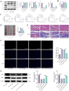
The protective effect of Sev on myocardial ischemia-reperfusion injury (I/R) in rats. (A) Serum cardiac troponin T levels; (B) Hemodynamic parameters, including heart rate (HR), left ventricular developed pressure (LVDP), maximal rates of left ventricular pressure increase (+dp/dtmax) and decrease (-dp/dtmax), rate-pressure product (RPP), and shock index (SI); (C) Myocardial infarct size measured by TTC staining; (D) Histological evaluation of myocardial tissue damage and fibrosis using HE and Masson staining. The scale bar is 100 μm; (E) TUNEL assay to determine the rate of myocardial cell apoptosis. The scale bar is 200 μm; (F) Prediction of the potential target USP7 of Sev using the SuperPred and Ubibrowser 2.0 databases; (G) RT-qPCR analysis of USP7 mRNA expression; (H) IHC detection of USP7 protein-positive expression changes. The scale bar is 100 μm. Data were analyzed using one-way ANOVA (A, C, E, G and H) or two-way ANOVA (B), followed by Tukey’s post-hoc test. N=10 (A and B), N=5 (CE and GH), ****p < 0.0001.
Figure 2
Figure 2
Overexpression of USP7 inhibits the protective effect of Sev on H9c2 cells under hypoxia/reoxygenation (H/R) injury. (A) RT-qPCR analysis of USP7 mRNA levels; (B) WB analysis of USP7 protein expression levels; (C) CCK-8 assay to evaluate cell viability; (D) LDH release assay to assess cell damage; (E) TUNEL assay to determine the proportion of apoptotic cells. The scale bar is 200 μm. Data were analyzed using one-way ANOVA followed by Tukey’s post-hoc test. N=5, ***p < 0.001, ****p < 0.0001.
Figure 3
Figure 3
Sev reduces oxidative stress and inhibits ferroptosis in H9c2 cells by suppressing USP7. (A) Levels of GSH and MDA to assess oxidative stress; (B) ROS levels to evaluate intracellular reactive oxygen species. The scale bar is 200 μm; (C) Fe²+ levels to measure intracellular iron content; (D) RT-qPCR analysis of mRNA expression of ferroptosis inhibitors GPX4 and FTH1; (E) WB analysis of GPX4 and FTH1 protein expression. Data are analyzed using one-way ANOVA followed by Tukey’s post hoc test. N=5, **p < 0.01, ***p < 0.001, ****p < 0.0001.
Figure 4
Figure 4
USP7 regulates PTEN stability through deubiquitination, contributing to the protective effects of Sev against ferroptosis. (A) Intersection of USP7 downstream targets predicted by the Ubibrowser database and ferroptosis-related targets identified from the Ferrdb database. (B) WB analysis of PTEN, PI3K, and AKT protein levels in myocardial tissues of I/R rats. (C) WB analysis of PTEN, PI3K, and AKT protein expression in H9c2 cells. (D) Co-IP assay confirming the interaction between USP7 and PTEN. (E) Analysis of PTEN ubiquitination levels following co-transfection with HA-Ub, Myc-PTEN, and Flag-USP7 plasmids. (F) Effect of USP7 knockdown on PTEN protein half-life after CHX treatment. Data were analyzed using one-way ANOVA (B and C) and two-way ANOVA (F), followed by Tukey’s post hoc test. N=5, ****p < 0.0001.
Figure 5
Figure 5
Knockdown of PTEN alleviates ferroptosis in H9c2 cells induced by USP7 overexpression through activation of the PI3K/Akt pathway. (A) RT-qPCR analysis of PTEN knockdown efficiency; (B) Western blot analysis of PTEN knockdown levels; (C) CCK-8 assay to assess cell viability; (D) LDH release assay to evaluate cellular damage; (E) TUNEL assay to measure apoptosis rates. The scale bar is 200 μm; (F) GSH and MDA assays to evaluate oxidative stress levels; (G) ROS detection assay. The scale bar is 200 μm; (H) Measurement of Fe²+ levels to assess intracellular iron content; (I) Western blot analysis of PI3K, AKT, GPX4, and FTH1 protein expression. Data were analyzed using one-way ANOVA followed by Tukey’s post hoc test. N=5, ***p < 0.001, ****p < 0.0001.
Figure 6
Figure 6
Validation of Sev’s protective effect against ferroptosis in cardiomyocytes through inhibition of USP7-mediated PTEN deubiquitination and activation of the PI3K/AKT signaling pathway in vivo. (A) Western blot analysis of USP7, PTEN, PI3K, and AKT protein expression in myocardial tissues from different treatment groups; (B) Serum cTnT levels; (C) Hemodynamic parameters, including heart rate (HR), stroke index (SI), left ventricular diastolic pressure (LVDP), +dp/dtmax, -dp/dtmax, and rate-pressure product (RPP); (D) Myocardial infarct size assessed by TTC staining; (E) Structural damage to cardiomyocytes observed by HE staining and. Myocardial fibrosis evaluated by Masson staining. The scale bar is 100 μm; (F) Cardiomyocyte apoptosis detected by TUNEL staining. The scale bar is 200 μm; (G) Expression levels of ferroptosis-related proteins GPX4 and FTH1 in myocardial tissue determined by Western blot analysis. Data were analyzed using one-way ANOVA (A, B, D, F and G) and two-way ANOVA (C), followed by Tukey’s post hoc test. N=10 (B and C), N=5 (A and DG), ns p > 0.05, ****p < 0.0001.

Similar articles

References

    1. Tian H, Zhao X, Zhang Y, Xia Z. Abnormalities of glucose and lipid metabolism in myocardial ischemia-reperfusion injury. Biomed Pharmacother. 2023;163:114827. doi: 10.1016/j.biopha.2023.114827 - DOI - PubMed
    1. Yi J, Zhu J, Wu J, Thompson CB, Jiang X. Oncogenic activation of PI3K-AKT-mTOR signaling suppresses ferroptosis via SREBP-mediated lipogenesis. Proc Natl Acad Sci U S A. 2020;117(49):31189–31197. doi: 10.1073/pnas.2017152117 - DOI - PMC - PubMed
    1. Zou R, Shi W, Qiu J, et al. Empagliflozin attenuates cardiac microvascular ischemia/reperfusion injury through improving mitochondrial homeostasis. Cardiovasc Diabetol. 2022;21(1):106. doi: 10.1186/s12933-022-01532-6 - DOI - PMC - PubMed
    1. Liu Y, Li X, Sun T, Li T, Li Q. Pyroptosis in myocardial ischemia/reperfusion and its therapeutic implications. Eur J Pharmacol. 2024;971:176464. doi: 10.1016/j.ejphar.2024.176464 - DOI - PubMed
    1. Qu Z, Pang X, Mei Z, et al. The positive feedback loop of the NAT10/Mybbp1a/p53 axis promotes cardiomyocyte ferroptosis to exacerbate cardiac I/R injury. Redox Biol. 2024;72:103145. doi: 10.1016/j.redox.2024.103145 - DOI - PMC - PubMed

MeSH terms

LinkOut - more resources