Case Reports
. 2025 Jul 19;17(7):e88305.
doi: 10.7759/cureus.88305. eCollection 2025 Jul.

Human Herpesvirus 6 Associated With a Secondary Central Nervous System Vasculitis in an Immunocompetent Adult: A Case Report

Affiliations
Case Reports

Human Herpesvirus 6 Associated With a Secondary Central Nervous System Vasculitis in an Immunocompetent Adult: A Case Report

Olivia Feuchter Ruy Sánchez et al. Cureus. .

Abstract

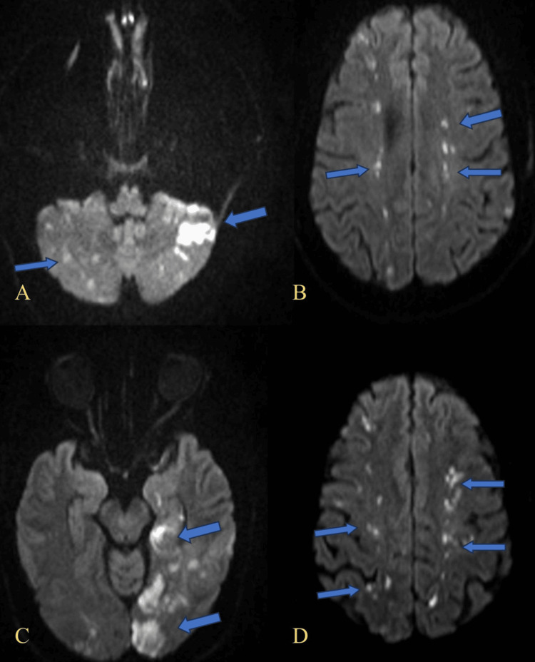
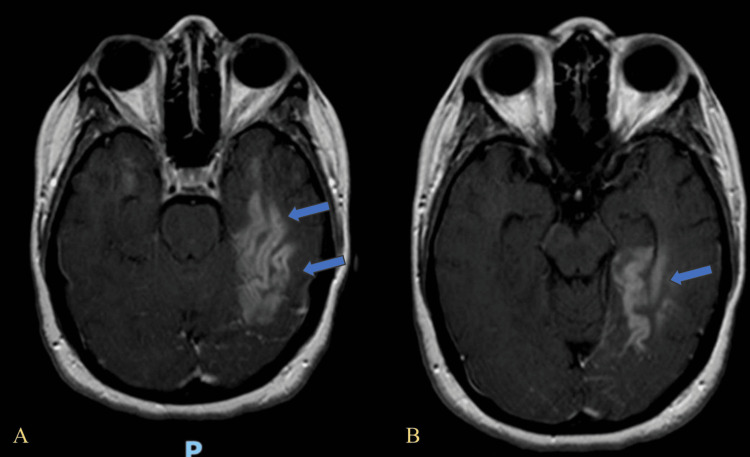
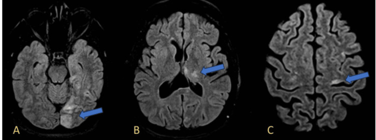
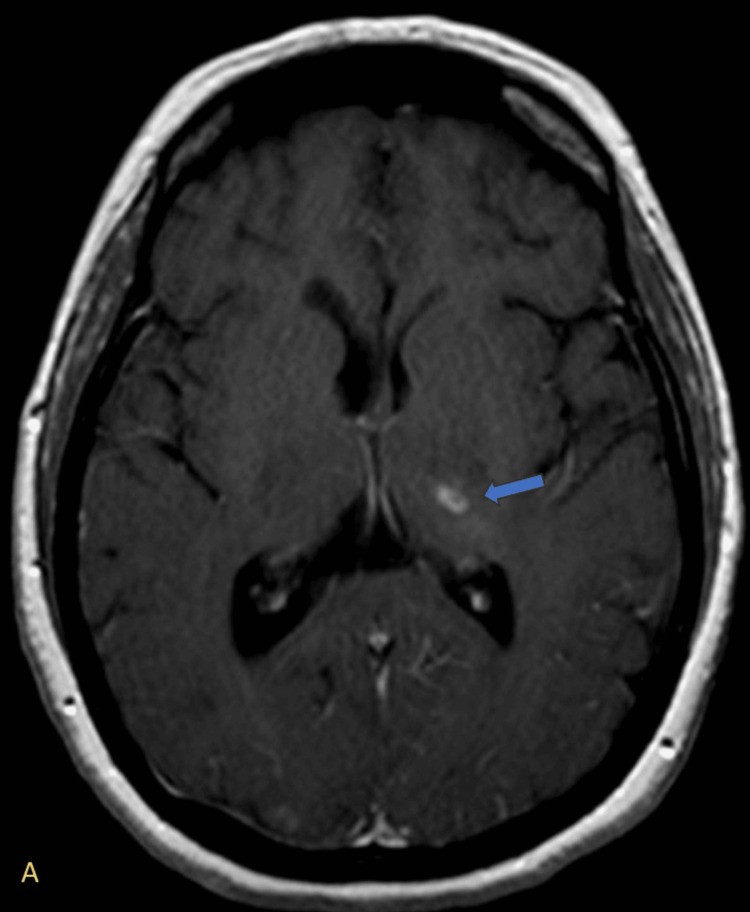
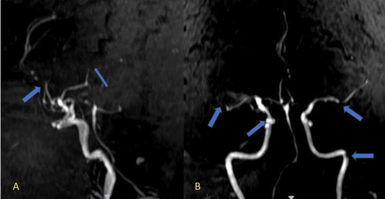
Human herpesvirus 6 (HHV-6) encephalitis typically affects children under three years of age, and adult cases are rare, usually occurring in immunocompromised individuals such as hematopoietic stem cell transplant recipients. HHV-6 can establish latency and reactivate under conditions of immunosuppression. Several neurotropic viruses, including varicella-zoster virus and herpes simplex virus, are known to induce central nervous system (CNS) vasculitis, but HHV-6 has not been clearly associated with this complication. We report the case of a 46-year-old immunocompetent woman presenting with encephalopathy, hemiparesis, and cerebellar signs. Brain MRI revealed ischemic lesions in multiple vascular territories and imaging features suggestive of cerebral vasculitis. Cerebrospinal fluid polymerase chain reaction (CSF PCR) was positive for HHV-6. Other etiologies, including neoplastic and autoimmune causes, were ruled out. The patient was treated with ganciclovir and dexamethasone at doses appropriate for CNS vasculitis. This case highlights a rare presentation of HHV-6-associated CNS vasculitis in an immunocompetent host and emphasizes the need to consider viral etiologies in the differential diagnosis of CNS vasculopathies.

Keywords: atypical neurological presentation; cerebrospinal fluid analysis; cns vasculitis; encephalitis; human herpesvirus 6; immunocompetent host; neuroimaging; pcr diagnostics; viral vasculopathy.


Conflict of interest statement

Human subjects: Informed consent for treatment and open access publication was obtained or waived by all participants in this study. Conflicts of interest: In compliance with the ICMJE uniform disclosure form, all authors declare the following: Payment/services info: All authors have declared that no financial support was received from any organization for the submitted work. Financial relationships: All authors have declared that they have no financial relationships at present or within the previous three years with any organizations that might have an interest in the submitted work. Other relationships: All authors have declared that there are no other relationships or activities that could appear to have influenced the submitted work.

Figures

Figure 1
Figure 1. Diffusion-weighted imaging (DWI) demonstrating multifocal areas of restricted diffusion. (A) Bilateral hyperintensities in the cerebellar hemispheres, more prominent on the left (blue arrows), compatible with acute ischemia. (B) Multiple foci of restricted diffusion in the bilateral subcortical white matter of the frontal lobes (blue arrows), indicative of acute embolic infarcts. (C) Hyperintense lesions in the left occipital lobe (blue arrows), consistent with acute ischemic injury. (D) Scattered foci of restricted diffusion in the bilateral parietal cortices (blue arrows), suggestive of multiple acute cortical infarctions.
DWI: diffusion-weighted imaging
Figure 2
Figure 2. Axial post-contrast T1-weighted MRI showing cortical and subcortical enhancement in the left temporal lobe. (A) Hyperintense signal involving the lateral aspect of the left temporal lobe cortex (blue arrows), consistent with cortical enhancement and encephalitic changes. (B) More medial axial slice showing confluent enhancement in the left mesial temporal lobe, including the hippocampus (blue arrow).
Figure 3
Figure 3. FLAIR sequences showing multifocal hyperintensities. (A) Hyperintense signal in the left occipital lobe (blue arrow), suggestive of acute infarction. (B) Restricted diffusion in the left thalamus (blue arrow), consistent with subcortical ischemia. (C) Cortical hyperintensity in the left frontal lobe, suggestive of acute cortical ischemia (blue arrow).
FLAIR: fluid-attenuated inversion recovery
Figure 4
Figure 4. Axial post-contrast T1-weighted MRI showing a ring-enhancing lesion in the left thalamus. (A) A small ring-enhancing lesion is noted in the left thalamus (blue arrow), characterized by peripheral contrast enhancement with a hypointense center. This radiologic pattern is suggestive of a ring-enhancing lesion, typically seen in infectious processes.
Figure 5
Figure 5. Magnetic resonance angiography (MRA) showing segmental narrowing of intracranial vessels. (A) MRA image reveals multifocal areas of segmental narrowing in the anterior cerebral artery (ACA) and middle cerebral artery (MCA) territories (blue arrows), suggestive of vasculitic changes. (B) Additional areas of irregular caliber and narrowing are observed in both anterior circulations and the basilar artery (blue arrows), supporting the presence of a diffuse vasculopathy.

References

    1. Human herpesvirus 6 encephalitis in immunocompetent and immunocompromised hosts. Berzero G, Campanini G, Vegezzi E, et al. Neurol Neuroimmunol Neuroinflamm. 2021;8 - PMC - PubMed
    1. Central nervous system vasculitis in adults: an update. Dutra LA, de Souza AW, Grinberg-Dias G, Barsottini OG, Appenzeller S. Autoimmun Rev. 2017;16:123–131. - PubMed
    1. Human herpesvirus 6 encephalitis. Gewurz BE, Marty FM, Baden LR, Katz JT. Curr Infect Dis Rep. 2008;10:292–299. - PubMed
    1. A rare case of HHV-6 encephalitis in an immunocompetent host: case report and literature review. Mekheal E, Tagliaferri AR, Vasquez KS, et al. Cureus. 2022;14:0. - PMC - PubMed

Publication types

LinkOut - more resources