

Navigating Ovarian-Adnexal Reporting and Data System Magnetic Resonance Imaging (O-RADS MRI): A Review of Its Evolution, Current Advances, and Persistent Challenges in Ovarian Imaging
- PMID: 40718198
- PMCID: PMC12291148
- DOI: 10.7759/cureus.86717
Navigating Ovarian-Adnexal Reporting and Data System Magnetic Resonance Imaging (O-RADS MRI): A Review of Its Evolution, Current Advances, and Persistent Challenges in Ovarian Imaging
Abstract
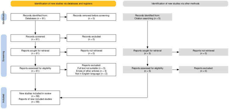
The Ovarian-Adnexal Reporting and Data System magnetic resonance imaging (O-RADS MRI) is a standardized risk stratification system designed to enhance uniform interpretation and reporting of adnexal masses on MRI. A PubMed search was conducted using the keyword "O-RADS MRI," yielding 61 articles in the search results. After excluding eight articles, 53 articles were selected. Additionally, five articles were identified through a citation search. A total of 58 articles were included in this literature review. Ultrasonography (USG) is the primary imaging modality used for evaluating adnexal lesions, with MRI reserved for cases that require further evaluation. Based on both USG and MRI imaging, various scores are assigned to a particular lesion. Contrast imaging, utilizing both ultrasound USG and MRI, is employed for the better characterization of lesions in terms of internal morphology, with a primary focus on solid components. Additionally, internal septation, wall characteristics, and other soft tissue components, including fat, fibrous tissue, and blood products, are also evaluated. Advanced MRI techniques, such as diffusion-weighted imaging and dynamic contrast-enhanced sequences, also help refine the final O-RADS score of a lesion. Contrast-Enhanced Computed Tomography (CECT) plays a predominant role in evaluating metastatic disease and has been established in five cases. This review article provides a comprehensive overview of O-RADS MRI, addressing its development, key imaging features, and practical application in a clinical setup. We discuss the diagnostic performance of O-RADS MRI in differentiating benign from malignant adnexal lesions, exploring its strengths in reducing inter-observer variability and guiding patient management. We also highlighted various comparative studies and trials that have shaped the evolution of the O-RADS MRI system over time. Furthermore, this article highlights the challenges associated with implementing O-RADS MRI, including potential pitfalls in interpretation, corroborations, and discordance with other imaging modalities, particularly USG, as well as the need for further validation studies.
Keywords: adnexal mass lesion; cect; dwi-adc; mri; o-rads mri; time-signal intensity curve; usg.
Copyright © 2025, Dev et al.
Conflict of interest statement
Conflicts of interest: In compliance with the ICMJE uniform disclosure form, all authors declare the following: Payment/services info: All authors have declared that no financial support was received from any organization for the submitted work. Financial relationships: All authors have declared that they have no financial relationships at present or within the previous three years with any organizations that might have an interest in the submitted work. Other relationships: All authors have declared that there are no other relationships or activities that could appear to have influenced the submitted work.
Figures
Similar articles
-
Systematic Review and Meta-Analysis of O-RADS Ultrasound and O-RADS MRI for Risk Assessment of Ovarian and Adnexal Lesions.AJR Am J Roentgenol. 2023 Jul;221(1):21-33. doi: 10.2214/AJR.22.28396. Epub 2023 Feb 1. AJR Am J Roentgenol. 2023. PMID: 36722758
-
Contrast-enhanced ultrasound using SonoVue® (sulphur hexafluoride microbubbles) compared with contrast-enhanced computed tomography and contrast-enhanced magnetic resonance imaging for the characterisation of focal liver lesions and detection of liver metastases: a systematic review and cost-effectiveness analysis.Health Technol Assess. 2013 Apr;17(16):1-243. doi: 10.3310/hta17160. Health Technol Assess. 2013. PMID: 23611316 Free PMC article.
-
The Role of Dynamic Contrast Enhanced Magnetic Resonance Imaging in Evaluating Prostate Adenocarcinoma: A Partially-Blinded Retrospective Study of a Prostatectomy Patient Cohort With Whole Gland Histopathology Correlation and Application of PI-RADS or TNM Staging.Prostate. 2025 Apr;85(5):413-423. doi: 10.1002/pros.24843. Epub 2024 Dec 19. Prostate. 2025. PMID: 39702937 Free PMC article.
-
MRI of pediatric ovarian masses: validation of the O-RADS framework.Eur Radiol. 2025 Aug;35(8):5073-5080. doi: 10.1007/s00330-025-11444-0. Epub 2025 Feb 13. Eur Radiol. 2025. PMID: 39939426 Free PMC article.
-
Magnetic resonance perfusion for differentiating low-grade from high-grade gliomas at first presentation.Cochrane Database Syst Rev. 2018 Jan 22;1(1):CD011551. doi: 10.1002/14651858.CD011551.pub2. Cochrane Database Syst Rev. 2018. PMID: 29357120 Free PMC article.
References
-
- Comparative study of the efficacy of the Ovarian-Adnexa Reporting and Data System ultrasound combined with contrast-enhanced ultrasound and the ADNEX MR scoring system in the diagnosis of adnexal masses. Wang T, Cui W, Nie F, et al. Ultrasound Med Biol. 2023;49:2072–2080. - PubMed
-
- Contemporary guidelines for adnexal mass imaging: a 2020 update. Stein EB, Roseland ME, Shampain KL, Wasnik AP, Maturen KE. Abdom Radiol (NY) 2021;46:2127–2139. - PubMed
-
- Accuracy and reproducibility of the O-RADS MRI risk stratification system based on enhanced non-DCE MRI in the assessment of adnexal masses. Wu M, Tang Q, Cai S, et al. Eur J Radiol. 2023;159:110670. - PubMed
Publication types
LinkOut - more resources
Full Text Sources