Urethral Leiomyoma in Pregnancy: A Rare Case of Symptomatic Pelvic Mass With Successful Surgical Management
- PMID: 40718265
- PMCID: PMC12291364
- DOI: 10.7759/cureus.86720
Urethral Leiomyoma in Pregnancy: A Rare Case of Symptomatic Pelvic Mass With Successful Surgical Management
Abstract
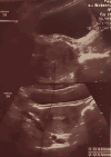
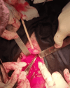
Leiomyomas, benign tumors derived from smooth muscle, are typically found in the uterus. Urethral involvement is an exceptionally rare occurrence, particularly in the context of a pre-fashioned thigh flap. Our case, therefore, presents a unique and intriguing instance, with only a few similar cases reported. We present a case of a 27-year-old primigravida at 11 weeks gestation with a protruding anterior vaginal mass, pelvic heaviness, and recurrent urinary complaints. A well-circumscribed vascular mass came out through the vulval orifice on physical examination. Transvaginal ultrasonography and pelvic magnetic resonance imaging revealed a 6×4 cm-sized mass that originated from the anterior vaginal wall, and it did not invade the adjacent structures. The surgical resection through anterior vaginal route with the use of spinal anesthesia. Histopathological examination revealed urethral leiomyoma, and the diagnosis was supported by positive immunoreactivity for the smooth muscle markers. The recovery following surgery was uneventful, and the pregnancy was carried to term with a successful normal delivery, instilling hope and optimism in similar cases. Our case underscores the crucial role of obstetricians and gynecologists in considering urethral leiomyoma in the differential diagnosis of anterior vaginal wall masses, even in pregnancy. This emphasis on early multimodal treatment can significantly impact the outcome; good results can be achieved, and pregnancy outcomes can be improved with curative therapy.
Keywords: fibroid uterus; leiomyoma; lower urinary tract symptoms; protruding vaginal mass; urethral leiomyoma; uterus like mass.
Copyright © 2025, Latoui et al.
Conflict of interest statement
Human subjects: Informed consent for treatment and open access publication was obtained or waived by all participants in this study. Conflicts of interest: In compliance with the ICMJE uniform disclosure form, all authors declare the following: Payment/services info: All authors have declared that no financial support was received from any organization for the submitted work. Financial relationships: All authors have declared that they have no financial relationships at present or within the previous three years with any organizations that might have an interest in the submitted work. Other relationships: All authors have declared that there are no other relationships or activities that could appear to have influenced the submitted work.
Figures


Similar articles
-
Surgical management of pelvic organ prolapse in women.Cochrane Database Syst Rev. 2013 Apr 30;(4):CD004014. doi: 10.1002/14651858.CD004014.pub5. Cochrane Database Syst Rev. 2013. Update in: Cochrane Database Syst Rev. 2016 Nov 30;11:CD004014. doi: 10.1002/14651858.CD004014.pub6. PMID: 23633316 Updated.
-
Pre-operative GnRH analogue therapy before hysterectomy or myomectomy for uterine fibroids.Cochrane Database Syst Rev. 2001;(2):CD000547. doi: 10.1002/14651858.CD000547. Cochrane Database Syst Rev. 2001. Update in: Cochrane Database Syst Rev. 2017 Nov 15;11:CD000547. doi: 10.1002/14651858.CD000547.pub2. PMID: 11405968 Updated.
-
Single-incision sling operations for urinary incontinence in women.Cochrane Database Syst Rev. 2017 Jul 26;7(7):CD008709. doi: 10.1002/14651858.CD008709.pub3. Cochrane Database Syst Rev. 2017. Update in: Cochrane Database Syst Rev. 2023 Oct 27;10:CD008709. doi: 10.1002/14651858.CD008709.pub4. PMID: 28746980 Free PMC article. Updated.
-
Use of endoanal ultrasound for reducing the risk of complications related to anal sphincter injury after vaginal birth.Cochrane Database Syst Rev. 2015 Oct 29;2015(10):CD010826. doi: 10.1002/14651858.CD010826.pub2. Cochrane Database Syst Rev. 2015. PMID: 26513224 Free PMC article.
-
Routine vaginal examinations for assessing progress of labour to improve outcomes for women and babies at term.Cochrane Database Syst Rev. 2013 Jul 15;(7):CD010088. doi: 10.1002/14651858.CD010088.pub2. Cochrane Database Syst Rev. 2013. Update in: Cochrane Database Syst Rev. 2022 Mar 4;3:CD010088. doi: 10.1002/14651858.CD010088.pub3. PMID: 23857468 Updated.
References
-
- Primary urethral leiomyoma in a female patient: a case report and review of the literature. Sharma R, Mitra SK, Dey RK, Basu S, Das RK. https://www.sciencedirect.com/science/article/pii/S1110570415000211 Afr J Urol. 2015;21:120–122.
-
- Laparoscopic management of a large urethral leiomyoma. Ciravolo G, Ferrari F, Zizioli V, Donarini P, Forte S, Sartori E, Odicino F. Int Urogynecol J. 2019;30:1211–1213. - PubMed
Publication types
LinkOut - more resources
Full Text Sources
