

Microscope-Integrated Indocyanine Green Videoangiography in Pediatric Spinal Dural Arteriovenous Fistula (SDAVF) Surgery: A Case Report on an Extremely Rare Vascular Malformation
- PMID: 40718324
- PMCID: PMC12296940
- DOI: 10.7759/cureus.86843
Microscope-Integrated Indocyanine Green Videoangiography in Pediatric Spinal Dural Arteriovenous Fistula (SDAVF) Surgery: A Case Report on an Extremely Rare Vascular Malformation
Abstract
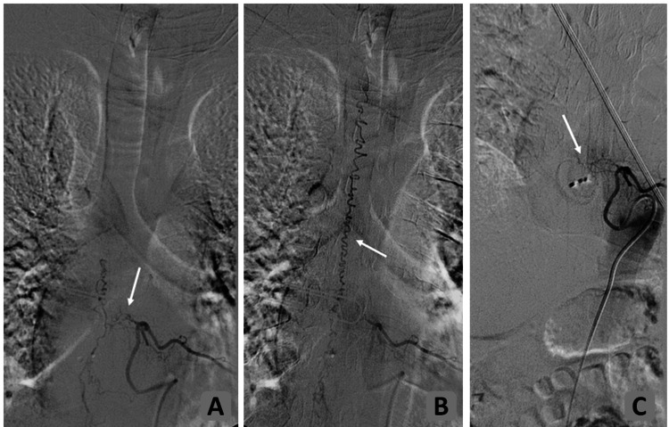
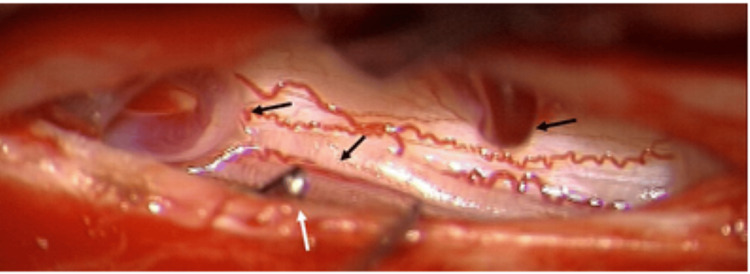
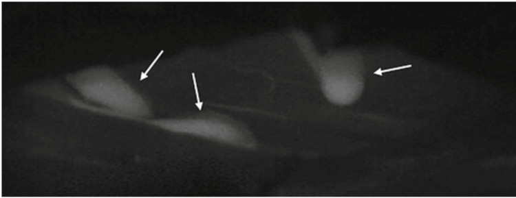
Spinal dural arteriovenous fistulas (SDAVFs) are exceptionally rare, acquired vascular malformations of the spinal cord, mostly affecting middle-aged to elderly men and typically presenting with progressive myelopathy. Given the diagnostic and therapeutic challenges of SDAVFs in children, we evaluated the feasibility and effectiveness of microscope-integrated indocyanine green (ICG) videoangiography as an intraoperative adjunct. While ICG use is well-documented in the surgical management of cranial aneurysms and adult spinal vascular malformations, its application in spinal arteriovenous malformations (AVM) in children remains limited and poorly characterized, and to date, its use in pediatric SDAVFs, especially, has not been reported. We present an exceptionally rare case of a pediatric SDAVF in a nine-year-old male patient who exhibited progressive, high-grade paraparesis and gait disturbances. The microsurgical disconnection of the fistula was performed at our institution. Intraoperative microscope-integrated ICG videoangiography was employed to localize the fistulous point at the Th7 level and to confirm the complete obliteration of the arteriovenous shunt in real time. No intraoperative or postoperative complications related to ICG administration were observed. Postoperative digital subtraction angiography (DSA) confirmed complete fistula closure. No recurrence or delayed complications were noted during a 70-month follow-up period. The results of this case report underscore the safety and effectiveness of intraoperative microscope-integrated ICG videoangiography for localizing and confirming the obliteration of pediatric SDAVF. This technique offers a promising alternative to routine postoperative DSA, reducing radiation exposure-associated risks in the pediatric population. Further studies on larger cohorts are warranted to validate these findings and standardize the use of ICG in pediatric spinal vascular surgery.
Keywords: intraoperative indocyanine green; microscope; pediatric; spinal angiography; spinal dural arteriovenous fistulas.
Copyright © 2025, Shabo et al.
Conflict of interest statement
Human subjects: Informed consent for treatment and open access publication was obtained or waived by all participants in this study. Conflicts of interest: In compliance with the ICMJE uniform disclosure form, all authors declare the following: Payment/services info: All authors have declared that no financial support was received from any organization for the submitted work. Financial relationships: All authors have declared that they have no financial relationships at present or within the previous three years with any organizations that might have an interest in the submitted work. Other relationships: All authors have declared that there are no other relationships or activities that could appear to have influenced the submitted work.
Figures

References
-
- Spinal epidural angiomatous malformations draining into intrathecal veins. Kendall BE, Logue V. Neuroradiology. 1977;13:181–189. - PubMed
-
- Intraspinal extramedullary arteriovenous fistulae draining into the medullary veins. Merland JJ, Riche MC, Chiras J. https://pubmed.ncbi.nlm.nih.gov/7276996/ J Neuroradiol. 1980;7:271–320. - PubMed
-
- [Spinal dural arteriovenous fistulas] (Article in German) Thron A. Radiologe. 2001;41:955–960. - PubMed
-
- Spinal dural arteriovenous fistulas: a congestive myelopathy that initially mimics a peripheral nerve disorder. Jellema K, Tijssen CC, van Gijn J. Brain. 2006;129:3150–3164. - PubMed
-
- Sacral origin of a spinal dural arteriovenous fistula: case report and review. Schaat TJ, Salzman KL, Stevens EA. Spine (Phila Pa 1976) 2002;27:893–897. - PubMed
Publication types
LinkOut - more resources
Full Text Sources