

In vitro models of muscle spindles: From traditional methods to 3D bioprinting strategies
- PMID: 40718504
- PMCID: PMC12290365
- DOI: 10.1177/20417314251343388
In vitro models of muscle spindles: From traditional methods to 3D bioprinting strategies
Abstract
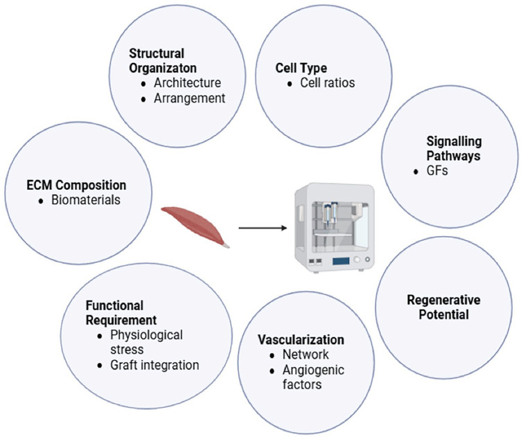
Muscle spindles are key proprioceptive mechanoreceptors composed of intrafusal fibres that regulate kinaesthetic sensations and reflex actions. Traumatic injuries and neuromuscular diseases can severely impair the proprioceptive feedback, yet the regenerative potential and cell-matrix interactions of muscle spindles remain poorly understood. There is a pressing need for robust tissue-engineered models to study spindle development, function and regeneration. Traditional approaches, while insightful, often lack physiological relevance and scalability. Three-dimensional (3D) bioprinting offers a promising approach to fabricate biomimetic, scalable, and animal-free muscle spindle constructs with controlled cellular architecture. Various bioprinting techniques - including inkjet, extrusion, digital light projection and laser-assisted bioprinting - have been explored for skeletal muscle fabrication, but replicating intrafusal fibre complexity remains a challenge. A major challenge lies in bioink development, where biocompatibility, printability and mechanical strength must be balanced to support intrafusal fibre differentiation and proprioceptive function. Recent molecular insights into spindle anatomy, innervation and extracellular matrix composition are shaping biofabrication strategies. This review discusses the current state of muscle spindle modelling, the application of 3D bioprinting in intrafusal fibre engineering, key challenges and future directions.
Keywords: bioink; bioprinting; hydrogel; intrafusal fibre; muscle spindle; proprioception; skeletal muscle; three-dimensional (3D) bioprinting; tissue engineering.
© The Author(s) 2025.
Conflict of interest statement
The author(s) declared no potential conflicts of interest with respect to the research, authorship, and/or publication of this article.
Figures


Similar articles
-
Macromolecular crowding-based biofabrication utilizing unmodified extracellular matrix bioinks.Acta Biomater. 2025 May 15;198:37-48. doi: 10.1016/j.actbio.2025.02.052. Epub 2025 Apr 22. Acta Biomater. 2025. PMID: 40268621
-
Iohexol as a refractive index tuning agent for bioinks in high cell density bioprinting.Biomater Sci. 2025 Jul 8;13(14):3958-3971. doi: 10.1039/d5bm00585j. Biomater Sci. 2025. PMID: 40512130
-
The Black Book of Psychotropic Dosing and Monitoring.Psychopharmacol Bull. 2024 Jul 8;54(3):8-59. Psychopharmacol Bull. 2024. PMID: 38993656 Free PMC article. Review.
-
Microfiber-Templated Porogel Bioinks Enable Tubular Interfaces and Microvascularization Down to the Building Blocks for 3D Bioprinting.Small. 2025 Jul;21(27):e2501594. doi: 10.1002/smll.202501594. Epub 2025 Mar 18. Small. 2025. PMID: 40099633
-
The Ethical Implications of Tissue Engineering for Regenerative Purposes: A Systematic Review.Tissue Eng Part B Rev. 2023 Apr;29(2):167-187. doi: 10.1089/ten.TEB.2022.0033. Epub 2022 Oct 20. Tissue Eng Part B Rev. 2023. PMID: 36112697 Free PMC article.
References
-
- Stillman BC. Making Sense of Proprioception: The meaning of proprioception, kinaesthesia and related terms. Physiotherapy 2002; 88: 667–676.
-
- Proske U, Gandevia SC. The proprioceptive senses: their roles in signaling body shape, body position and movement, and muscle force. Physiol Rev 2012; 92: 1651–1697. - PubMed
Publication types
LinkOut - more resources
Full Text Sources
