. 2025 Jul 11:13:1577669.
doi: 10.3389/fcell.2025.1577669. eCollection 2025.

CPT1A mediated preservation of mitochondrial inhibits pyroptosis in pancreatic acinar cells

Affiliations

CPT1A mediated preservation of mitochondrial inhibits pyroptosis in pancreatic acinar cells

Yijiang Liu et al. Front Cell Dev Biol. .

Abstract

Introduction: Carnitine palmitoyltransferase 1A (CPT1A) is crucial for mitochondrial function, and its dysfunction has been linked to the development of several diseases. However, the role of CPT1A in severe acute pancreatitis (SAP) and its underlying mechanisms remain unclear. Mitochondrial damage-mediated pyroptosis has been identified as a critical factor in pancreatic acinar cell death during SAP. this study aimed to evaluate the protective role of CPT1A in SAP and investigate its association with pancreatic acinar cell pyroptosis.

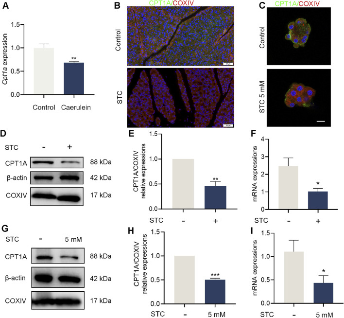
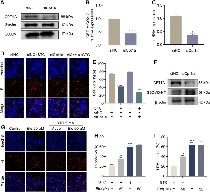
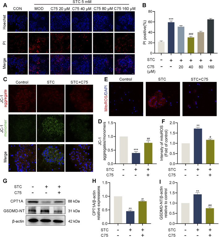
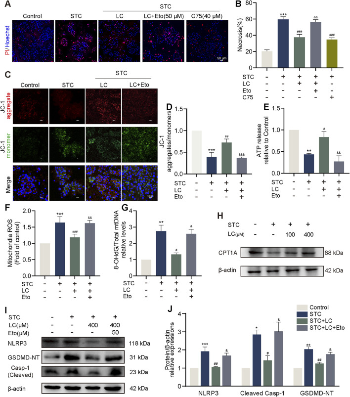
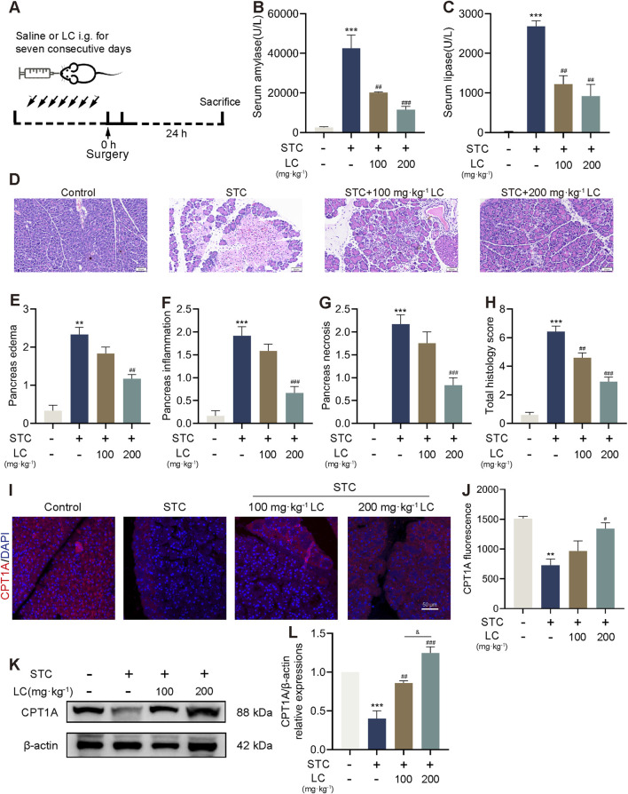
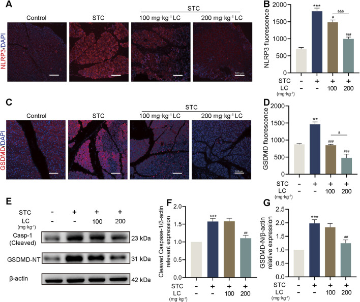
Methods: SAP models were established in male C57BL/6 mice by retrograde injection of 3% sodium taurocholate (STC) into the pancreatic duct and in primary acinar cells treated with 5 mM STC. Changes in Cpt1a mRNA and protein expression were assessed. Pancreatic pyroptosis was evaluated via activation of NLRP3 inflammasome-related proteins. Cpt1a was knocked down (siRNA) or inhibited (etomoxir) in cells. Cell viability was measured using Hoechst/PI staining, western blotting, and LDH release assays. The effects of CPT1A activators (C75, L-carnitine(LC)) on mitochondrial function (ΔΨm, mtROS, ox-mtDNA release) were examined in acinar cells.

Results: In STC-induced SAP models (in vivo and in vitro), CPT1A expression was downregulated. Activating CPT1A with C75 or LC protected mitochondrial function (preserving ΔΨm, reducing mtROS, inhibiting ox-mtDNA release), thereby suppressing pyroptosis. LC treatment alleviated SAP in mice by inhibiting the NLRP3/GSDMD/Caspase-1 pathway and reducing acinar cell pyroptosis.

Discussion: These findings reveal a novel protective mechanism of CPT1A in SAP. Enhancing CPT1A activity preserves mitochondrial functions and suppresses NLRP3/GSDMD-mediated pancreatic acinar cell pyroptosis, highlighting CPT1A as a potential therapeutic target.

Keywords: CPT1A; L-carnitine; acute pancreatitis; mitochondrial dysfunction; pyroptosis.


Conflict of interest statement

The authors declare that the research was conducted in the absence of any commercial or financial relationships that could be construed as a potential conflict of interest.

Figures

FIGURE 1
FIGURE 1
Pancreatic CPT1A was downregulated in STC-induced SAP mice. (A) Analysis of Cpt1a gene expression in dataset GSE3644. (B) Immunofluorescence staining of CPT1A in the pancreas of mice. Scale bar: 50 μm. (C) CPT1A fluorescence was observed in STC-treated primary acinar cells. Scale bar: 20 μm. (D,E) Protein levels of CPT1A, COXIV and β-actin in STC-treated pancreas tissue of mice. (F) Cpt1a mRNA expression in the pancreas of mice. (G,H) Protein levels of CPT1A, COXIV and β-actin in primary acinar cells in response to STC (5 mM). (I) Cpt1a mRNA expression in primary acinar cells. All data are presented as means ± SEM, n = 3–5. *p < 0.05, **p < 0.01, ***p < 0.001 vs. Control group. STC: sodium taurocholate.
FIGURE 2
FIGURE 2
Inhibition of CPT1A exacerbates STC-induced cell death. Inhibition of CPT1A exacerbates STC-induced cell death. (A,B) Protein levels of CPT1A, COXIV and β-actin in 266-6 cells in response to siNC and siCpt1a. (C) Cpt1a mRNA relative expressions to control. (D) Representative images of Hoechst 33342 (blue) and PI (red) staining of 266-6 cells treated with STC, STC+siCpt1a, respectively. Scale bar: 50 μm. (E) CCK8 was used to determine the cell viability. (F) Protein levels of CPT1A and GSDMD-NT in 266-6 cells received different treatments. (G) Representative images of Hoechst 33342 (blue) and PI (red) staining of primary acinar cells received different treatments. Scale bar: 50 μm. (H) The ratio of PI positive cells in different group. (I) LDH release in the lysate of acinar cells. All data are presented as means ± SEM, n = 3–5. *p < 0.05, **p < 0.01, ***p < 0.001 vs. Control group or siNC group. ### p < 0.001 vs. STC+siNC group. NC: negative control; STC: sodium taurocholate; Eto: Etomoxir.
FIGURE 3
FIGURE 3
CPT1A attenuates STC-induced cell pyroptosis through preserving mitochondrial function. CPT1A attenuates STC-induced cell pyroptosis through preserving mitochondrial function. (A,B) Representative images and quantification of Hoechst 33342 (blue) and PI (red) staining of primary acinar cells received different concentrations of C75 treatments. (C,D) JC-1 fluorescent staining of primary acinar cells in different treatment groups. Scale bar: 50 μm. (E,F) Fluorescent quantification and imaging of mtROS in primary acinar cells in different treatment groups. (G–I) Protein levels of CPT1A and GSDMD-NT in the primary acinar cell received different treatments. β-actin served as the loading control (n = 3). All data are presented as means ± SEM, n = 3–5. *p < 0.05, **p < 0.01, ***p < 0.001 vs. Control group. # p < 0.05, ## p < 0.01, ### p < 0.001 vs. STC group.
FIGURE 4
FIGURE 4
LC protects against STC-induced acinar cell pyroptosis via CPT1A. (A,B) Representative images and quantification of Hoechst 33342 (blue) and PI (red) staining of primary acinar cells received different treatments. Scale bar: 50 μm. (C,D) JC-1 fluorescent staining of primary acinar cells in different treatment groups. Scale bar: 50 μm. (E) The release of ATP levels in the supernatant of cultured primary acinar cells received different treatments. (F) Quantification of mtROS in primary acinar cells in different treatment groups. (G) The ratio of 8-OHdG to total mtDNA content in primary acinar cells received different treatments. (H) Protein levels of CPT1A and β-actin in the primary acinar cells received different treatments. (I,J) Protein levels of NLRP3, cleaved Caspase-1 and GSDMD-NT in the primary acinar cell received different treatments. β-actin served as the loading control (n = 3). Data are presented as means ± SEM, n = 3–5. *p < 0.05, **p < 0.01, ***p < 0.001 vs. Control group. # p < 0.05, ## p < 0.01, ### p < 0.001, vs. STC group. & p < 0.05, && p < 0.01, &&& p < 0.001 vs. STC+LC treatment group. STC: sodium taurocholate; LC: L-carnitine; Eto: Etomoxir.
FIGURE 5
FIGURE 5
CPT1A activation ameliorates STC-induced pancreatic acinar cells injury. (A) The scheme for LC pretreatment of mice with SAP. (B,C) Serum amylase and lipase levels. (D) Representative H&E images. (E–H) Histological scores of the pancreas (edema, inflammatory infiltration, necrosis and total scores). (I,J) Immunofluorescence staining of CPT1A in the pancreas of mice received different treatments. Scale bar: 100 μm. (K,L) Protein levels of CPT1A and β-actin in the pancreas of mice received different treatments. All data are presented as means ± SEM, n = 3–5. **p < 0.01, ***p < 0.001 vs. Control group. # p < 0.05, ## p < 0.01, ### p < 0.001 vs. STC group.
FIGURE 6
FIGURE 6
CPT1A protects against SAP in mice by inhibiting the NLRP3/GSDMD-mediated pyroptosis signalling pathway. (A–D) Immunofluorescence staining of NLRP3 and GSDMD in the pancreas of mice received different treatments. Scale bar: 100 μm. (E–G) Protein levels of cleaved Caspase-1 and GSDMD-NT in the pancreas of mice received different treatments. β-actin served as the loading control. All data are presented as means ± SEM, n = 3–5. *p < 0.05, **p < 0.01, ***p < 0.001 vs. Control group. # p < 0.05, ## p < 0.01, ### p < 0.001 vs. STC group. & p < 0.05, &&& p < 0.001 vs. LC (100 mg/kg) treatment group.

Similar articles

References

    1. Adeva-Andany M. M., Calvo-Castro I., Fernandez-Fernandez C., Donapetry-Garcia C., Pedre-Pineiro A. M. (2017). Significance of l-carnitine for human health. Iubmb Life 69 (8), 578–594. 10.1002/iub.1646 - DOI - PubMed
    1. Aho H. J., Koskensalo S. M. L., Nevalainen T. J. (1980). Experimental pancreatitis in the rat. Sodium taurocholate-induced acute haemorrhagic pancreatitis. Scand. J. Gastroenterology 15 (4), 411–416. 10.3109/00365528009181493 - DOI - PubMed
    1. Al Mamun A., Suchi S. A., Aziz M. A., Zaeem M., Munir F., Wu Y. Q., et al. (2022). Pyroptosis in acute pancreatitis and its therapeutic regulation. Apoptosis 27 (7-8), 465–481. 10.1007/s10495-022-01729-w - DOI - PubMed
    1. Al Mamun A., Wu Y. Q., Monalisa I., Jia C., Zhou K. L., Munir F., et al. (2021). Role of pyroptosis in spinal cord injury and its therapeutic implications. J. Adv. Res. 28, 97–109. 10.1016/j.jare.2020.08.004 - DOI - PMC - PubMed
    1. Baron T. H., DiMaio C. J., Wang A. Y., Morgan K. A. (2020). American gastroenterological association clinical practice update: management of pancreatic necrosis. Gastroenterology 158 (1), 67–75. 10.1053/j.gastro.2019.07.064 - DOI - PubMed

LinkOut - more resources