. 2025 Sep 1;329(3):C754-C767.
doi: 10.1152/ajpcell.00249.2025. Epub 2025 Jul 28.

Ablation of UCP-1+ cells impacts FAP dynamics in muscle regeneration

Affiliations

Ablation of UCP-1+ cells impacts FAP dynamics in muscle regeneration

Jacob C Parson et al. Am J Physiol Cell Physiol. .

Abstract

Uncoupling protein-1 (UCP-1+) cells found in brown adipose tissue and subtypes of white (a.k.a. beige) adipose tissue have been a focus of intensive investigation for their role in energy metabolism and are emerging as potential endocrine regulators of physiology. More recently, UCP-1+ subpopulations have also been found in skeletal muscle fibro-adipogenic progenitors (FAPs), which play an important role in regeneration. Both UCP-1+ adipocytes and FAPs secrete promyogenic cytokines further supporting their potential for proregenerative signaling. To investigate whether signaling from UCP-1+ cells does indeed promote regeneration, we examined injury-induced muscle regeneration in a mouse model with constitutive UCP-1+ cell ablation (UCP1-DTA) at three time points: early [3 and 7 days post injury (dpi)], intermediate (14 dpi), and late (21 dpi). We hypothesized that without UCP-1+ cells, muscle regeneration would be impaired at all time points. At 3 and 7 dpi, we found significantly reduced numbers of FAPs in male UCP1-DTA mice, but with no accompanying changes in muscle-derived stem (satellite) cells or immune cells. However, at 14 dpi, we observed significantly higher numbers of FAP in male UCP1-DTA mice and evidence of ongoing early-phase regeneration, including significantly increased histological and gene expression of early regenerative markers and significantly smaller regenerating fibers. However, these changes were not associated with fibrosis and fatty infiltration typical of impaired regeneration, nor were differences in contractile force recovery observed between genotypes. These findings suggest that UCP-1+ cells (adipocytes or FAPs) may regulate FAP dynamics in early regeneration, but without major effects on the recovery of structure and function.NEW & NOTEWORTHY Accumulating evidence suggests that UCP-1+ brown and beige adipose tissue impact muscle metabolism and that UCP-1+ FAPs impact atrophy, fibrosis, and fatty infiltration in a chronic injury model. This is the first report to examine muscle regeneration in the absence of brown fat and to explore the loss (rather than the addition) of UCP-1+ FAPs. We find that loss of both UCP-1+ adipocytes and FAPs only mildly impacts muscle regeneration, without disturbance of structural or functional recovery.

Keywords: brown adipose tissue; contractile function; fibro-adipogenic progenitors; glycerol injury.


Figures

Figure 1.
Figure 1.. Fibro-adipogenic progenitor (FAP), but not muscle-derived stem cell (MuSC), dynamics are affected by UCP-1+ cell ablation in male mice following glycerol-induced injury.
(A) Representative identification of FAP (green) and MuSC (pink) populations by flow cytometry. (B-C) Quantification of FAP and MuSC populations at 3 and 7 days post injury (dpi) in male and female WT and UCP1-DTA tibialis anterior muscle as a percentage of CD45− cells (parent). (D) Representative image of the FAP marker platelet-derived growth factor receptor alpha (Pdgfrα; green) immunostaining in histological sections at 14dpi counterstained with laminin (gray), and DAPI (blue). (E) Quantification of Pdgrα+ area fraction over the entire muscle section, indicative of area fraction occupied by FAPs (F) Gene expression of Pdgrα (Pdgfra) relative to GAPDH and normalized to the saline treated littermate control group. (G) Representative image of the MuSC marker paired box 7 (Pax7; pink) immunostaining in histological sections at 14dpi with laminin (gray), and DAPI (blue). (H) Quantification of Pax7+ nuclei/mm2, over the entire muscle section, indicative of MuSC density. (I) Gene expression of Pax7 (Pax7) relative to GAPDH and normalized to the saline treated littermate control group. Statistical analyses: N=5-8 mice per group. (B-C) 3-way ANOVA, Šídák’s multiple comparisons test; (E-F; H-I) 2-way ANOVA, Šídák’s multiple comparisons test.
Figure 2.
Figure 2.. Immune cell populations dynamics are not affected by UCP-1+ cell ablation, but are affected by sex following glycerol-induced injury.
(A) Representative identification of CD11b+/F4/80+ macrophages (red), F4/80-CD11b+/Ly6G− monocytes (burgundy), and F4/80−/CD11b+/Ly6G+ neutrophils (orange) by flow cytometry. (B-D) Quantification of macrophage, monocyte and neutrophil populations at 3 and 7 days post injury (dpi) in male and female WT and UCP1-DTA tibialis anterior muscle as a percentage of single cells. (E) Representative image of the pan-macrophage marker CD68 (red) and type 2 macrophage marker CD206 (green) immunostaining in histological sections at 14dpi counterstained with DAPI (blue). (F-G) Quantification of type 1 (CD68+/CD206−) and type 2 (CD68+/CD206+) macrophages per mm2, over the entire muscle section, indicative of macrophage density. Statistical analyses: N=5-8 mice per group. (B-D) 3-way ANOVA, Šídák’s multiple comparisons test; (F-G) 2-way ANOVA, Šídák’s multiple comparisons test. * p<0.05, ** p<0.01
Figure 3.
Figure 3.. UCP1-DTA Mice Show Signs of Delayed Regeneration but similar structural pathology at 14dpi following glycerol-induced injury.
(A) Representative identification of regenerating myofibers marked by embryonic myosin heavy chain (EmbMHC; red) immunostaining in histological sections of tibialis anterior muscle with laminin (gray), and DAPI (blue). (B) Quantification of EmbMHC area fraction over the entire muscle section, indicative of area fraction occupied by newly regenerated fibers. (C) Quantification of the average cross-sectional area of centrally-nucleated fibers over the entire muscle section, indicative of the average size of regenerated fibers. (D-E) Gene expression of regenerating markers EmbMHC (Myh3) and myogenin (Myog) relative to GAPDH and normalized to the saline treated littermate control group. (F) Representative identification of intramuscular adipose tissue (IMAT) by oil red O staining of histological sections. (G) Quantification of IMAT area fraction over the entire muscle section. (H) Gene expression of adiponectin (Adipoq) relative to GAPDH and normalized to the saline treated littermate control group. (I) Representative identification of collagen in the extracellular matrix by picrosirius red staining of histological sections. (J) Quantification of Sirius Red fibrosis area fraction over the entire muscle section (K) Gene expression of collagen 1 alpha 1 chain (Col1a1) relative to GAPDH and normalized to the saline treated littermate control group. Statistical analyses: N=5-7 mice per group. (B-E; G-H; J-K) 2-way ANOVA, Šídák’s multiple comparisons test. * p<0.05.
Figure 4.
Figure 4.. Association of FAPs with IMAT, fibrosis, and newly regenerated fibers varies between genotypes.
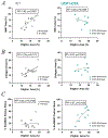
Linear regression of Pdgfrα+ area fraction against IMAT (A), Collagen (B) and EmbMHC (C) area fractions in WT (left) and UCP1-DTA (right) quantified from histological sections at 14dpi (data in Fig 3). Open circles indicate female and closed circles male data points. Data was combined for both sexes for the regression analysis. Statistical analyses: N=10-14 mice per genotype, simple linear regression.
Figure 5.
Figure 5.. UCP1-DTA mice recover contractile tension following GLY-induced injury similarly to WT mice.
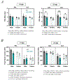
(A) 5th toe extensor digitorum longus (EDL) masses at 14 and 21 days post injury (dpi) in male and female WT and UCP1-DTA mice. (B) Peak tetanic tension (tetanic force normalized to PCSA) at 14 and 21 days post injury (dpi) in male and female WT and UCP1-DTA mice. Statistical analyses: N=5-8 mice per group. 3-way ANOVA, within-subjects matching (GLY-contralateral SAL) Šídák’s multiple comparisons test. * p<0.05, *** p<0.005

References

    1. Van Thi-Tuong Nguyen, Van Vu V, and Van Pham P Brown adipocyte and browning thermogenesis: metabolic crosstalk beyond mitochondrial limits and physiological impacts. Adipocyte 12: 2237164, 2023. - PMC - PubMed
    1. Carpentier AC, Blondin DP, Haman F, and Richard D. Brown Adipose Tissue-A Translational Perspective. Endocr Rev 44: 143–192, 2023. - PMC - PubMed
    1. Loncar D Convertible adipose tissue in mice. Cell Tissue Res 266: 149–161, 1991. - PubMed
    1. Shabalina IG, Petrovic N, de Jong JM, Kalinovich AV, Cannon B, and Nedergaard J. UCP1 in brite/beige adipose tissue mitochondria is functionally thermogenic. Cell Rep 5: 1196–1203, 2013. - PubMed
    1. Ahmad B, Vohra MS, Saleemi MA, Serpell CJ, Fong IL, and Wong EH. Brown/Beige adipose tissues and the emerging role of their secretory factors in improving metabolic health: The batokines. Biochimie 184: 26–39, 2021. - PubMed

LinkOut - more resources