Comparative Study
. 2025 Jul 28;7(8):e1292.
doi: 10.1097/CCE.0000000000001292. eCollection 2025 Aug 1.

Comparative Evaluation of Lipopolysaccharide Administration Methods to Induce Acute Lung Injury in Murine Models: Efficacy, Consistency, and Technical Considerations

Affiliations
Comparative Study

Comparative Evaluation of Lipopolysaccharide Administration Methods to Induce Acute Lung Injury in Murine Models: Efficacy, Consistency, and Technical Considerations

Eva Kuhar et al. Crit Care Explor. .

Abstract

Context: Direct preclinical lipopolysaccharide acute lung injury (ALI) models are commonly used to study acute respiratory distress syndrome. Differences in lipopolysaccharide delivery methods may impact lung injury severity and reproducibility.

Hypothesis: We hypothesized that the severity and variability of ALI outcomes in mice would differ depending on the technique of lipopolysaccharide administration.

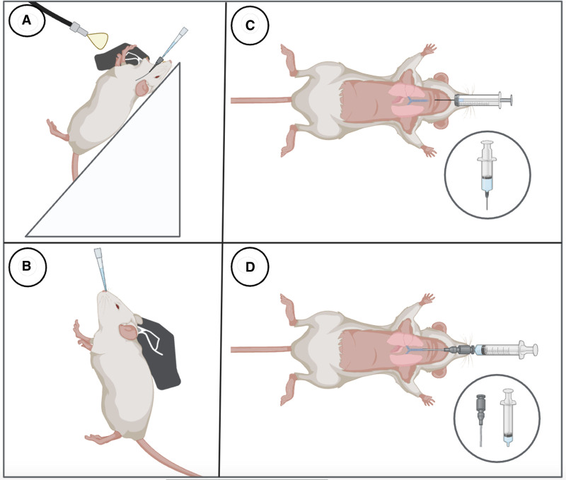
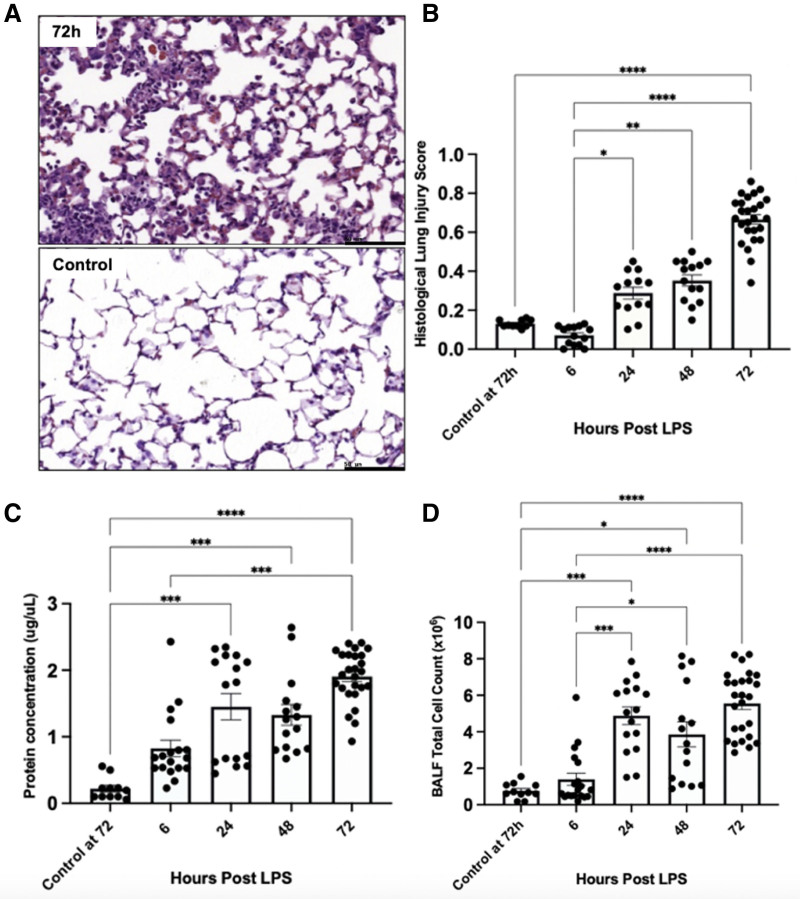
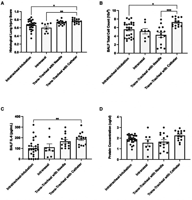
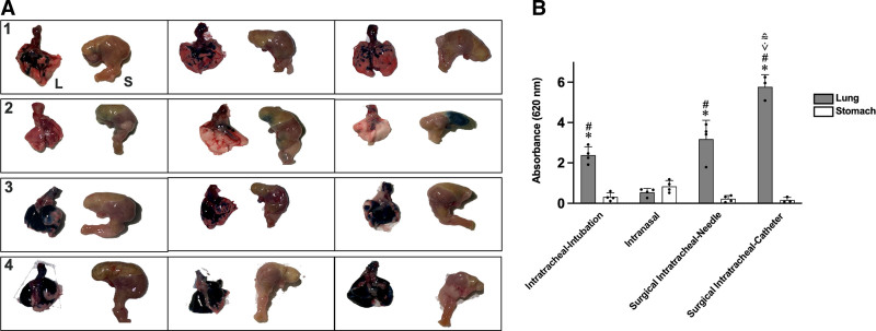
Methods and models: Male and female C57BL/6 mice were administered lipopolysaccharide (2.25 mg/kg) via four methods: 1) intratracheal intubation; 2) intranasal; 3) surgical transtracheal by either needle puncture; or 4) by catheter. ALI severity and variability were assessed at 72 hours post-lipopolysaccharide via histological scoring and bronchoalveolar lavage fluid (BALF) analysis (total protein, cell counts, interleukin-6 [IL-6]). The relative distribution of Evans Blue dye was also assessed for each model (lungs vs. stomach).

Results: Distinct lung injury patterns were observed between the four methods. The transtracheal with catheter method demonstrated significantly greater lung injury scores than the intratracheal intubation and intranasal techniques. Both transtracheal methods produced greater alveolar neutrophil counts, increased proteinaceous debris, fewer hyaline membranes, and lower variability than non-surgical techniques. The transtracheal with catheter method produced higher BALF total cell counts and IL-6 levels than intratracheal intubation. Transtracheal methods also resulted in more localized Evans Blue dye distribution in the lungs. Male mice exhibited more severe lung injury scores and higher BALF protein concentrations than females.

Interpretation and conclusions: This study demonstrates that the choice of technique to administer lipopolysaccharide impacts injury severity, phenotype, and variability. The surgical transtracheal with catheter technique produced the most robust and least variable ALI phenotype; however, this technique is associated with increased procedural complexity. Our results will allow researchers to tailor their model choice to align with their specific study objectives.

Keywords: acute lung injury; acute respiratory distress syndrome; biological sex; disease models; lipopolysaccharide; sex factors.


Conflict of interest statement

Ms. Kuhar was supported by a Canada Graduate Scholarship with the Canadian Institutes of Health Research (CIHR). Dr. Lalu wassupported by the Ottawa Hospital Anesthesia Alternate Funds Association, the Canadian Anesthesiologists’ Society Career Investigator Award, and a University of Ottawa Junior Research Chair in Innovative Translational Research. Dr. Zhang holds a Robert and Dorothy Pitts Chair in Acute Care and Emergency Medicine, a joint Hospital-University Endowed Chair between the University of Toronto, Unity Health Toronto, and the St. Michael’s Hospital Foundation. Mr. Jeffers is supported by the CIHR and Vanier Canada Graduate Scholarship. The remaining authors have disclosed that they do not have any potential conflicts of interest.

Figures

Figure 1.
Figure 1.
Diagram of four techniques of lipopolysaccharide administration. The administration techniques are as follows: A, intratracheal instillation-intubation; B, intranasal instillation; C, transtracheal instillation with needle; D, transtracheal instillation with catheter. The diagram was created using BioRender.
Figure 2.
Figure 2.
Time-course assessment of inflammatory and lung injury outcomes following lipopolysaccharide (LPS) administration via intratracheal intubation. Representative lung section from the time-course: A, 72 hr post-LPS administration; B, histological lung injury score; C, protein concentration in bronchoalveolar lavage fluid (BALF); and D, BALF total cell count were measured at different time points following LPS administration via the intratracheal intubation technique to assess lung injury progression. Control groups were included for baseline comparisons. Lung tissue sections were stained with hematoxylin and eosin and scored, with results expressed as mean ± sem (n = 10–15 per group). Total protein concentration in BALF was determined using the bicinchoninic acid assay (BCA) and presented as mean ± sem (n = 10–15 per group). BALF total cell count, indicating cell influx, was measured using a Cell Countess and reported as mean ± sem (n = 10–15 per group). Statistical analyses were conducted using the Kruskal-Wallis test followed by Dunn’s post hoc test, with p < 0.05 considered statistically significant. Significance is indicated as follows: *p < 0.05, **p < 0.01, ***p < 0.001, and ****p < 0.0001.
Figure 3.
Figure 3.
Comparison of four lipopolysaccharide (LPS) administration techniques on outcomes of acute lung injury: Histological evidence of tissue injury, inflammatory markers, and alveolar permeability. A, Histological lung injury score, B, bronchoalveolar lavage fluid (BALF) total cell count, C, interleukin-6 (IL-6) activity in BALF, and D, protein concentration in BALF were measured in mice 72 hr after LPS administration via four different techniques: intratracheal intubation, intranasal, transtracheal with needle, and transtracheal with catheter. Lung tissue sections were stained with hematoxylin and eosin and evaluated under a light microscope for histological scoring, with scores presented as mean ± sem (n = 8–15 per group). BALF total cell count, an indicator of cell influx, was assessed using a Cell Countess and presented as mean ± sem (n = 8–15 per group). Levels of the proinflammatory cytokine IL-6 in BALF were quantified using enzyme-linked immunosorbent assay, with results shown as mean ± sem (n = 8–15 per group). Total protein concentration in BALF was measured using the bicinchoninic acid assay and expressed as mean ± sem (n = 8–15 per group). Statistical analyses were performed using the Kruskal-Wallis test followed by Dunn’s post hoc test to compare differences among groups, with p < 0.05 considered statistically significant. Significance is indicated as follows: *p < 0.05, **p < 0.01, ***p < 0.001, and ****p < 0.0001.
Figure 4.
Figure 4.
Evans blue dye distribution and quantification in different lipopolysaccharide administration techniques. Representative images (n = 3 mice per technique) show the distribution of Evans Blue dye in the lungs and stomach 15 min after administration via four different techniques: A1, intratracheal intubation, A2, intranasal, A3, transtracheal with needle, and A4, transtracheal with catheter. The images illustrate the distribution patterns specific to each administration technique. B, Quantification of Evans Blue dye in lung and stomach tissues for each administration techniques. Dye concentration was assessed by measuring absorbance at 620 nm, providing an evaluation of tissue distribution. Results are presented as mean ± sem (n = 3– 4 per group). Statistical analyses were conducted using two-way analysis of variance followed by Tukey post hoc test to compare differences among groups. Statistical significance is indicated as follows: *Indicates a significant difference compared with the stomach tissue within the same technique. #Indicates a significant difference compared with the lung tissue of the intranasal instillation group. v̇Indicates a significant difference compared with the lung tissue of the intratracheal intubation group. ≅Indicates a significant difference compared with the lung tissue of the surgical intratracheal needle group.

Similar articles

References

    1. Bellani G, Laffey JG, Pham T, et al. ; LUNG SAFE Investigators: Epidemiology, patterns of care, and mortality for patients with acute respiratory distress syndrome in intensive care units in 50 countries. JAMA 2016; 315:788–800 - PubMed
    1. Rubenfeld GD, Caldwell E, Peabody E, et al. : Incidence and outcomes of acute lung injury. N Engl J Med 2005; 353:1685–1693 - PubMed
    1. Matute-Bello G, Frevert CW, Martin TR: Animal models of acute lung injury. Am J Physiol Lung Cell Mol Physiol 2008; 295:L379–L399 - PMC - PubMed
    1. Mizgerd JP, Skerrett SJ: Animal models of human pneumonia. Am J Physiol Lung Cell Mol Physiol 2008; 294:L387–L398 - PubMed
    1. Matute-Bello G, Downey G, Moore BB, et al. ; Acute Lung Injury in Animals Study Group: An official American Thoracic Society Workshop Report: Features and measurements of experimental acute lung injury in animals. Am J Respir Cell Mol Biol 2011; 44:725–738 - PMC - PubMed

Publication types

Substances

LinkOut - more resources