. 2025 Jul 28;21(7):e1012881.
doi: 10.1371/journal.ppat.1012881. eCollection 2025 Jul.

Targeting translation initiation yields fast-killing therapeutics against the zoonotic parasite Cryptosporidium parvum

Affiliations

Targeting translation initiation yields fast-killing therapeutics against the zoonotic parasite Cryptosporidium parvum

Meng Li et al. PLoS Pathog. .

Abstract

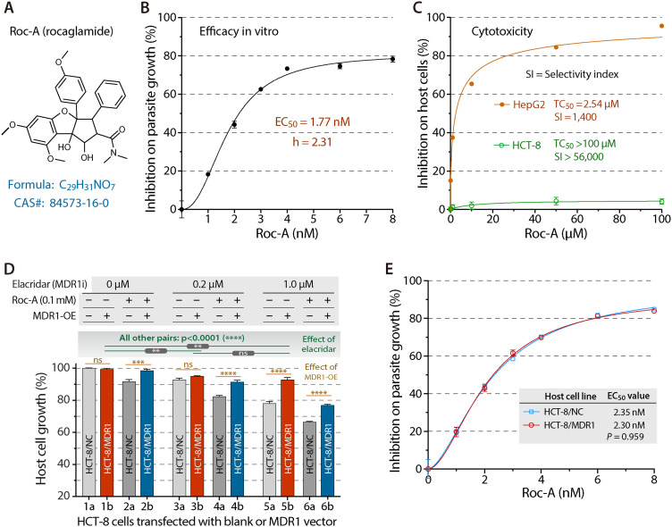
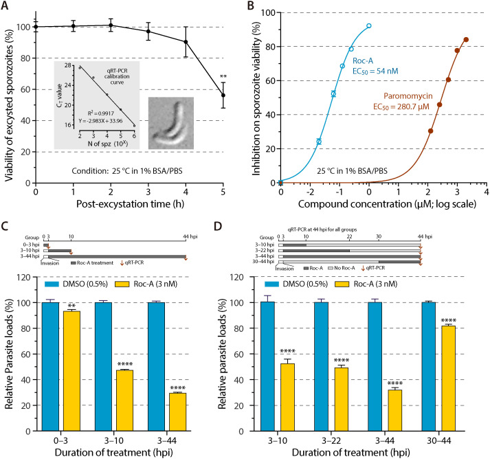
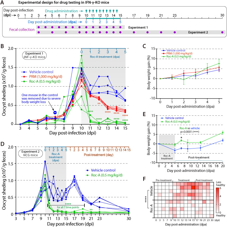
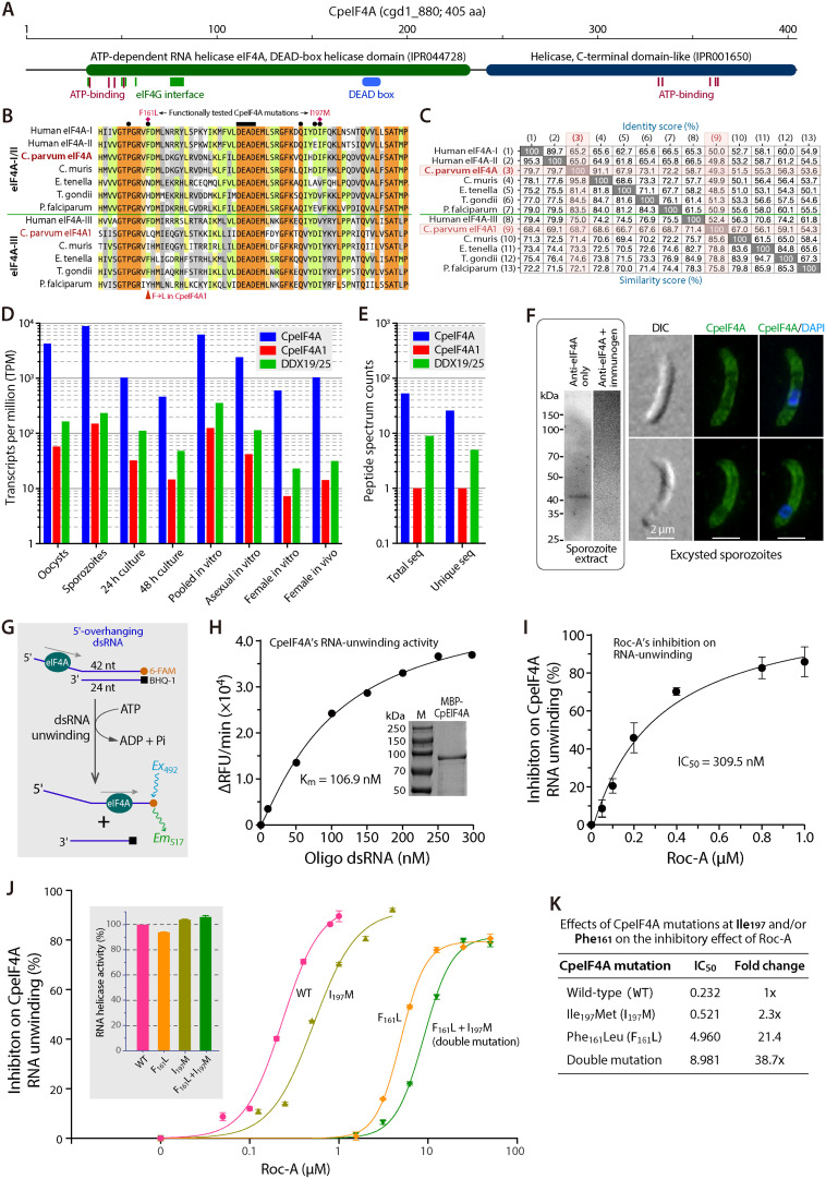
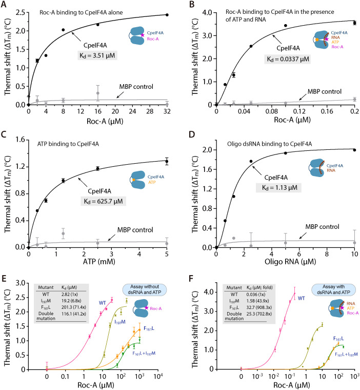
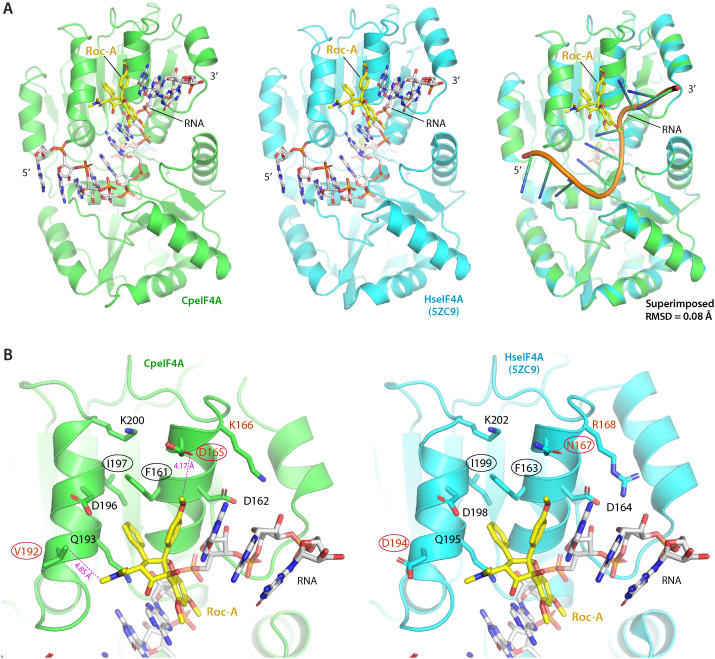
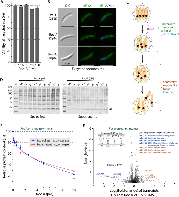
Cryptosporidium parvum is a zoonotic apicomplexan that causes moderate-to-severe watery diarrhea in children, immunocompromised patients, and neonatal ruminants, yet no fully effective drug is available. We show that the parasite's eukaryotic initiation factor 4A (CpeIF4A; a DEAD-box RNA helicase in the eIF4F translation-initiation complex) can be exploited as a fast-killing therapeutic target. Rocaglamide A (Roc-A), a plant-derived rocaglate, binds the CpeIF4A-RNA-ATP complex with high affinity (Kd = 33.7 nM) and blocks protein synthesis in excysting sporozoites (IC50 ≈ 3.7 µM). In host-cell culture, Roc-A suppresses intracellular growth with nanomolar potency (EC50 = 1.77 nM) and a selectivity index exceeding 56,000 in HCT-8 cells and 1,400 in HepG2 cells. A five-day oral regimen (0.5 mg/kg/d) reduced oocyst shedding by >90% within 48 h in interferon-γ-knockout mice and by 70-90% from day 2 onward without rebound during a 15-day follow-up in NCG mice. Two amino-acid differences at the Roc-A binding surface (D165 and V192 in CpeIF4A vs. N167 and D194 in the human ortholog) offer a foothold for medicinal optimization toward greater parasite selectivity. These findings establish translation initiation as an unexplored but tractable pathway for anti-cryptosporidial drug discovery and position Roc-A as a promising lead compound.


Conflict of interest statement

The authors have declared that no competing interests exist.

Figures

Fig 1
Fig 1. Rocaglamide A (Roc-A) selectively inhibits C. parvum growth by acting on the parasite eIF4A target.
(A) Chemical structure of Roc-A (CAS 84573-16-0), a highly selective inhibitor of eukaryotic initiation factor 4A (eIF4A). (B) Dose-response curve for Roc-A in a 44-h infection assay: C. parvum was grown in HCT-8 monolayers and parasite burden quantified by qRT-PCR targeting 18S rRNA (Cp18S). EC50 = 1.77 nM. (C) Host-cell cytotoxicity. HCT-8 and HepG2 cells were exposed to Roc-A for 44 h and viability measured by MTS. Selectivity index (SI) = TC50/ EC50. (D) Confirmation that Roc-A is an MDR1 substrate. HCT-8 cells transiently over-expressing human MDR1 (HCT-8/MDR1) or vector control (HCT-8/NC) were treated with 0.1 µM Roc-A in the absence or presence of the MDR1 inhibitor elacridar. MDR1 over-expression increased host-cell tolerance to Roc-A in every elacridar group. Significance by Tukey’s multiple comparisons: ** p < 0.01, *** p < 0.001, **** p < 0.0001; ns, not significant. (E) Over-expression of MDR1 does not alter Roc-A efficacy against C. parvum: inhibitory curves and EC50 values are identical in HCT-8/MDR1 and HCT-8/NC monolayers, demonstrating that the antiparasitic effect is on-target within the parasite.
Fig 2
Fig 2. Rocaglamide A (Roc-A) reduces sporozoite viability and blocks invasion and early intracellular development of C. parvum.
(A) Time-dependent viability of free sporozoites held at 25°C in PBS (pH 7.4) containing 1% BSA. Viability was measured by qRT-PCR for C. parvum 18S rRNA (Cp18S). Insets: calibration curve (CT vs. sporozoite number) and representative phase-contrast image of two excysted sporozoites. (B) Viability of excysted sporozoites after 2 h exposure to Roc-A or paromomycin (PMM) at the indicated concentrations (1% BSA/PBS, 25°C), quantified by Cp18S qRT-PCR. (C) Effect of Roc-A (3 nM in 0.5% DMSO; ~ EC63) during invasion (0–3 hpi) or first-generation merogony (3–10 hpi) compared with continuous treatment (3–44 hpi). Parasite burden was measured by qRT-PCR at the end of each exposure; schematic timeline is shown above the graph. (D) Drug-withdrawal assay distinguishing parasiticidal from parasitistatic action. Following invasion, cultures received Roc-A (3 nM) for 3–10 hpi or 3–22 hpi; drug was then removed and parasites were allowed to develop until 44 hpi. A continuous 3–44 hpi treatment served as reference; timeline is illustrated above the graph. For panels C and D, oocysts were added to HCT-8 monolayers, allowed to excyst and invade for 3 h, and uninvaded parasites were washed away before the indicated treatments. Statistical significance was determined by Sidak’s multiple comparisons test (** p < 0.01, **** p < 0.0001 versus the 0.5% DMSO control at the same time point).
Fig 3
Fig 3. Rocaglamide A (Roc-A) is highly efficacious in two murine models of chronic C. parvum infection.
(A) Schematic of the dosing protocol: IFN-γ-knockout (IFN-γ-KO) or NCG mice were infected orally with 5 × 104 oocysts, allowed to establish infection for 10 days, and then treated twice daily for 5 days with Roc-A (0.5 mg/kg/d), paromomycin (PRM, 1,000 mg/kg/d; IFN-γ-KO study only), or vehicle (1% DMSO). (B, C) IFN-γ-KO study (n = 5 per group). Roc-A reduced oocyst shedding by >90% within 24 h of the first dose and sustained suppression through day 5 (B), while body-weight trends were comparable to vehicle and PRM groups (C). One vehicle-treated mouse was removed on day 3 owing to excessive weight loss. (D–F) NCG study (n = 5 per group). Roc-A again produced a rapid, ≥ 90% decline in oocyst shedding that persisted without rebound through the 15-day follow-up (D); treated mice gained more weight (E) and had higher composite health scores (F) than vehicle controls. Oocyst output was quantified by qPCR, calibrated against standard curves generated by spiking known oocyst numbers into negative feces and processing identically. Statistical differences versus vehicle were assessed by Tukey’s multiple comparisons test (** p < 0.01; **** p < 0.0001). Error bars denote SEM.
Fig 4
Fig 4. Molecular and biochemical characterization of C. parvum eIF4A (CpeIF4A).
(A) Domain architecture of CpeIF4A (cgd1_880; 405 aa) predicted by InterProScan, showing N-terminal and C-terminal helicase domains, ATP-binding motifs, and eIF4G-contact residues. (B) Alignment of the conserved DEAD-box region from CpeIF4A, human eIF4A-I/II, orthologs from C. muris, E. tenella, T. gondii, and P. falciparum, and the eIF4A-III–type proteins (CpeIF4A1, human eIF4A-III, and their apicomplexan orthologs). The bar marks the canonical DEAD motif; filled circles denote Roc-A contact residues (PDB 5ZC9). Red diamonds indicate positions mutated in this study. (C) Pairwise identity and similarity scores for the sequences in panel B. (D, E) Relative abundance of CpeIF4A versus CpeIF4A1 across parasite stages, extracted from CryptoDB transcriptomes (D) and proteomes (E); CpeIF4A is expressed ≥25-fold higher at every stage (see S1–S2 Tables). (F) Detection of native CpeIF4A. Left, western blot of sporozoite lysate probed with an affinity-purified rabbit anti-CpeIF4A peptide antibody; a single band appears at the expected size. Right, immunofluorescence shows cytoplasmic localization with stronger signal beneath the plasma membrane (green); nuclei are counter-stained with DAPI (blue). DIC, differential-interference-contrast image. (G–I) RNA-unwinding assay for recombinant MBP-CpeIF4A (scheme in G). Michaelis–Menten plot for duplex RNA (H; inset: SDS-PAGE of purified protein) and dose–response curve for Roc-A inhibition (I). (J) Effect of point mutations Phe161Leu (F161L), Ile197Met (I197M), and the double mutant on helicase activity (inset) and binding affinity to Roc-A. (K) Corresponding shifts in Roc-A sensitivity: IC50 increases 2.3-fold (I197M), 21.4-fold (F161L), and 38.7-fold (double mutant), indicating Phe161 is the predominant determinant of high-affinity binding.
Fig 5
Fig 5. Rocaglamide A (Roc-A) binds CpeIF4A with highest affinity when duplex RNA and ATP are present.
(A–B) Thermal-shift assay (TSA) of Roc-A binding to apo-CpeIF4A (A) and to CpeIF4A pre-incubated with duplex RNA and ATP (B). The substrates tighten the interaction, lowering the apparent Kd from 3.51 µM to 33.7 nM (≈102-fold increase in affinity). (C, D) Control TSA curves for CpeIF4A binding to the individual substrates ATP (C) and duplex RNA (D). (E, F) Effect of point mutations F161L, I197M, and the double mutant (F161L + I197M) on Roc-A affinity in the absence (E) or presence (F) of RNA + ATP. I197M raises the Kd ~ 7-fold, F161L ~ 71-fold, and the double mutant ~41-fold in the apo state. When RNA + ATP are included, the losses deepen to ~44-, ~ 908-, and ~703-fold, respectively, confirming that Phe161 is the principal determinant of high-affinity binding.
Fig 6
Fig 6. Structural comparison of Roc-A binding to C. parvum eIF4A (CpeIF4A) and human eIF4A (HseIF4A).
(A) Homology model of CpeIF4A (left) built on the crystal structure of the HseIF4A–AMP-PNP–Roc-A–polypurine RNA complex (center; PDB 5ZC9). Right, superposition of the two structures illustrates their overall similarity. (B) Close-up of the Roc-A–binding pocket in CpeIF4A (left) and HseIF4A (right). Residues essential for ligand contact are labeled; conserved positions are in black, parasite-specific substitutions in red. Black-circled residues (Phe161, Ile197) were validated by mutagenesis, whereas the red-circled substitutions (Asp165, Val192) represent sites that could be exploited to enhance Roc-A selectivity for the parasite enzyme.
Fig 7
Fig 7. Rocaglamide A (Roc-A) suppresses protein synthesis but not global transcription in excysting C. parvum sporozoites.
(A) Viability of sporozoites released in vitro for 2 h in the presence of 0–100 µM Roc-A. Cp18S qRT-PCR shows no loss of viability at any concentration tested. (B) Morphology and CpeIF4A localization after 2 h exposure to 5 or 10 µM Roc-A. Sporozoites were stained with affinity-purified anti-CpeIF4A (green) and DAPI (blue); no changes are evident. (C) Schematic of excystation. Roc-A can reach sporozoites only after the oocyst suture ruptures and parasites exit. (D) Western blot of non-secreted (pellet) and secreted (supernatant) proteins from sporozoites excysted for 2 h with increasing Roc-A. A pan-Cryptosporidium antibody detects dose-dependent loss of most pellet bands, whereas only a single 28-kDa secreted band (arrowhead) declines. (E) Quantification of band intensity versus Roc-A concentration. Blue, total non-secreted proteins; red, 28-kDa secreted protein. Error bars, SEM. (F) Volcano plot of RNA-seq data from sporozoites treated for 2 h with 0.15 µM Roc-A (~EC70) versus 0.5% DMSO. Of 3,783 transcripts detected, none meet the stringent threshold |log2FC| > 2, q < 0.05; only seven low-abundance, uncharacterized genes change under the relaxed criterion |log2FC| > 2, p < 0.05.

Similar articles

References

    1. Ryan UM, Feng Y, Fayer R, Xiao L. Taxonomy and molecular epidemiology of Cryptosporidium and Giardia - a 50 year perspective (1971-2021 ). Int J Parasitol. 2021;51(13-14):1099–119. doi: 10.1016/j.ijpara.2021.08.007 - DOI - PubMed
    1. Ryan U, Zahedi A, Paparini A. Cryptosporidium in humans and animals-a one health approach to prophylaxis. Parasite Immunol. 2016;38(9):535–47. doi: 10.1111/pim.12350 - DOI - PubMed
    1. Checkley W, White AC Jr, Jaganath D, Arrowood MJ, Chalmers RM, Chen X-M, et al. A review of the global burden, novel diagnostics, therapeutics, and vaccine targets for cryptosporidium. Lancet Infect Dis. 2015;15(1):85–94. doi: 10.1016/S1473-3099(14)70772-8 - DOI - PMC - PubMed
    1. Innes EA, Chalmers RM, Wells B, Pawlowic MC. A one health approach to tackle Cryptosporidiosis. Trends Parasitol. 2020;36(3):290–303. doi: 10.1016/j.pt.2019.12.016 - DOI - PMC - PubMed
    1. Mmbaga BT, Houpt ER. Cryptosporidium and Giardia infections in children: a review. Pediatr Clin North Am. 2017;64(4):837–50. doi: 10.1016/j.pcl.2017.03.014 - DOI - PubMed

MeSH terms

Substances