Review
. 2025 Aug 12;19(31):28021-28109.
doi: 10.1021/acsnano.5c00706. Epub 2025 Jul 28.

Extracellular Vesicles for Clinical Diagnostics: From Bulk Measurements to Single-Vesicle Analysis

Affiliations
Review

Extracellular Vesicles for Clinical Diagnostics: From Bulk Measurements to Single-Vesicle Analysis

Hai Linh Tran et al. ACS Nano. .

Abstract

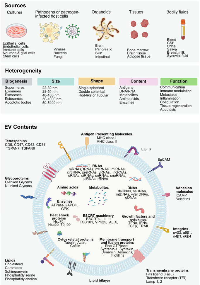
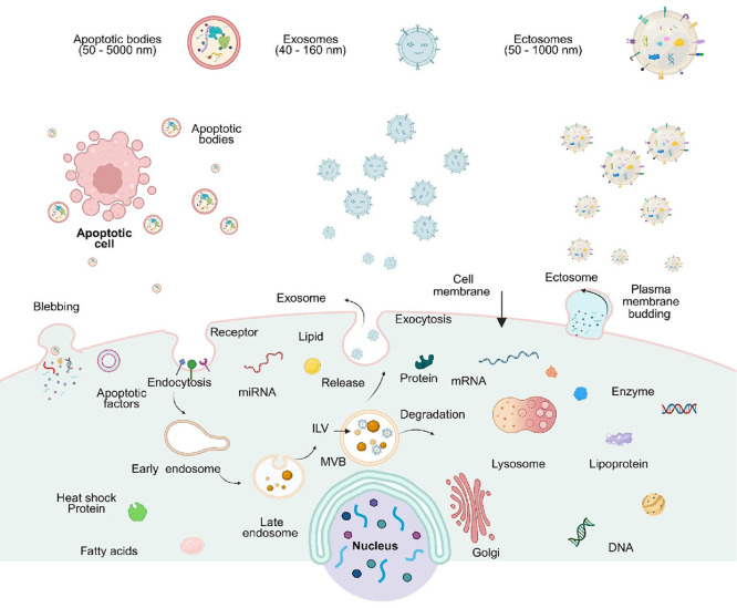
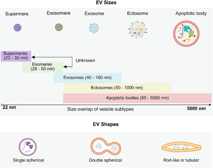
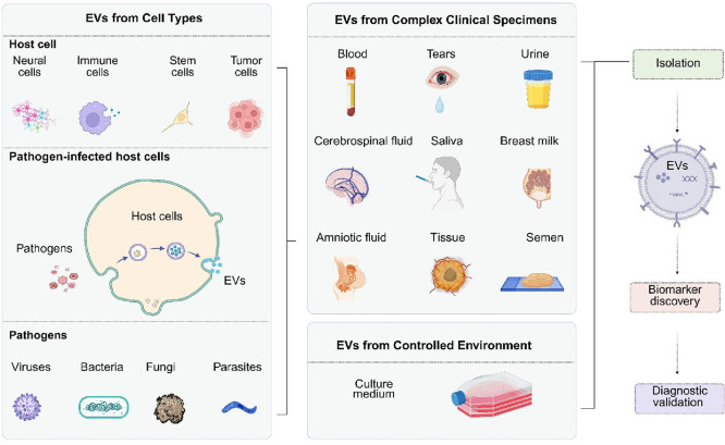
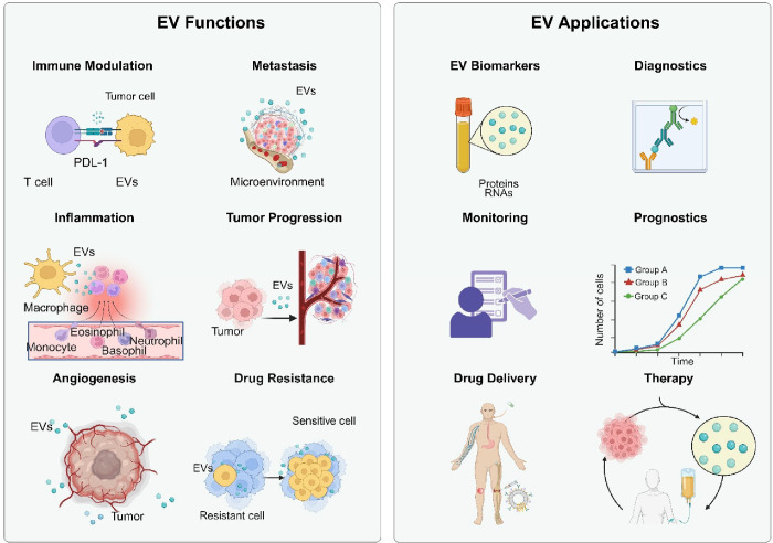
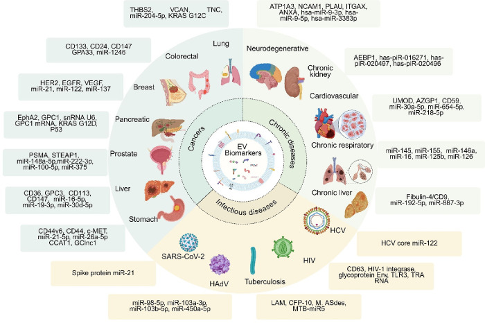
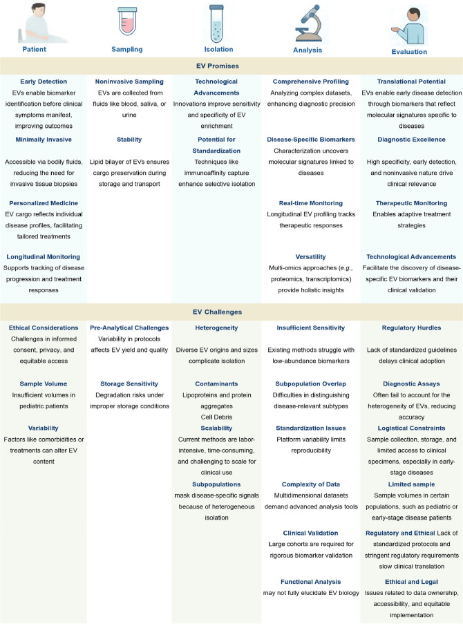
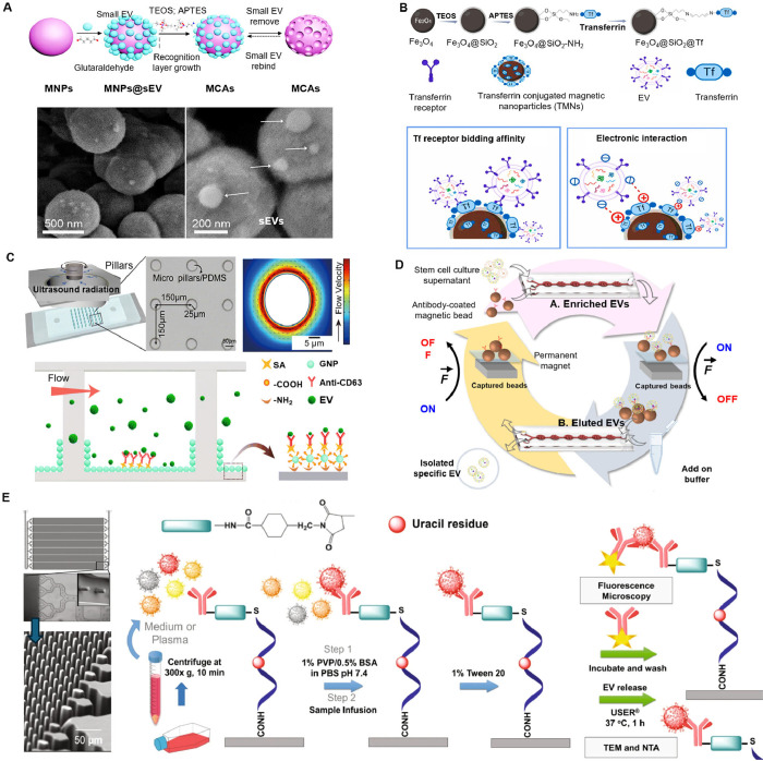
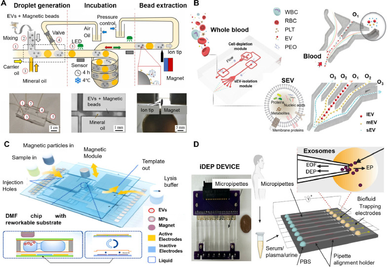
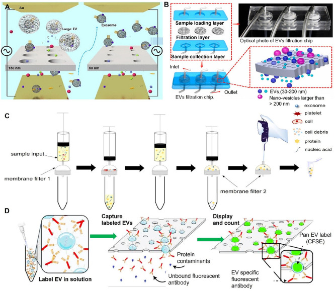
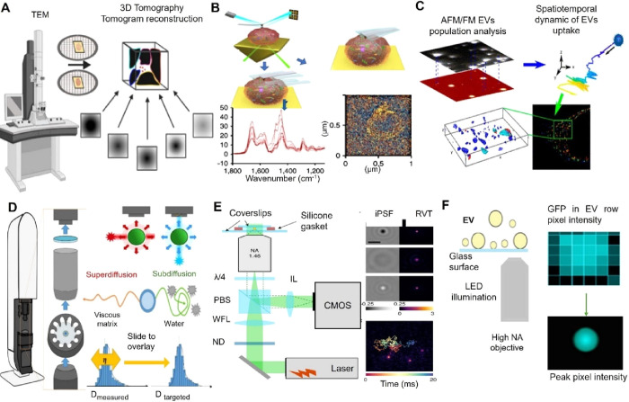
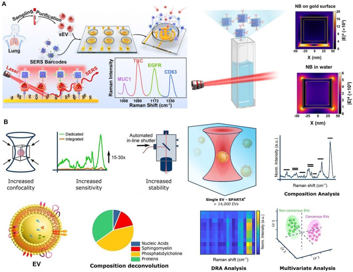
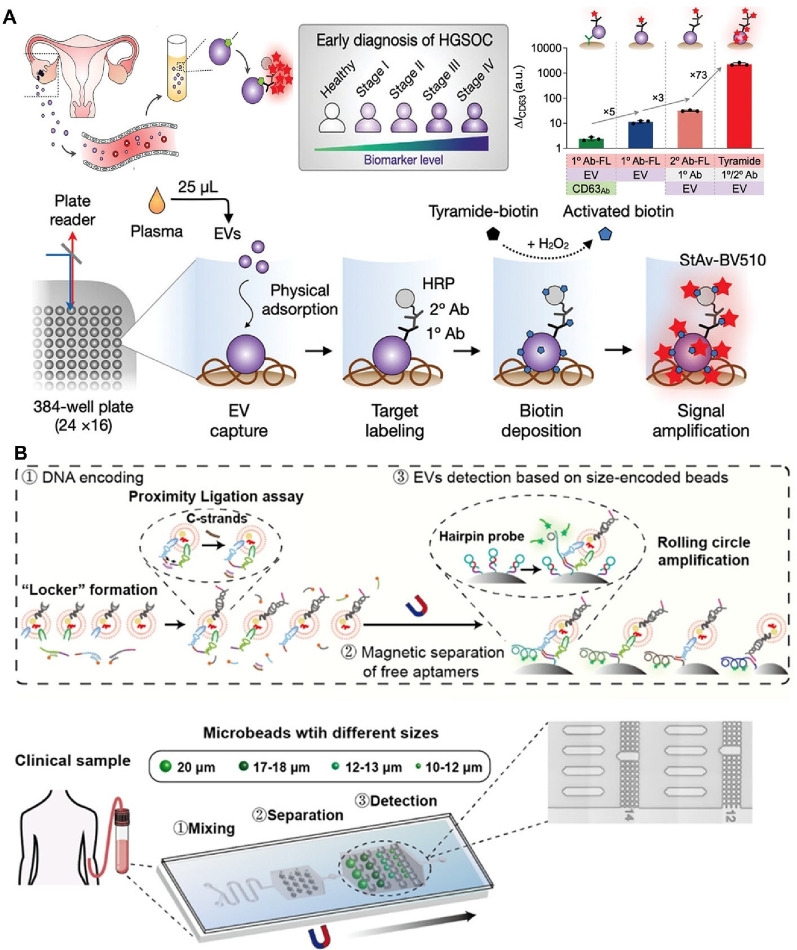
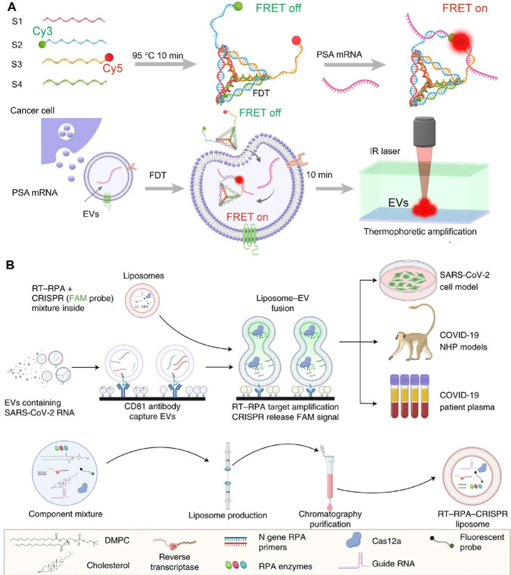
Extracellular vesicles (EVs) play a crucial role in intercellular communication, signaling pathways, and disease pathogenesis by transporting biomolecules such as DNA, RNA, proteins, and lipids derived from their cells of origin, and they have demonstrated substantial potential in clinical applications. Their clinical significance underscores the need for sensitive methods to fully harness their diagnostic potential. In this comprehensive review, we explore EV heterogeneity related to biogenesis, structure, content, origin, sample type, and function roles; the use of EVs as disease biomarkers; and the evolving landscape of EV measurement for clinical diagnostics, highlighting the progression from bulk measurement to single vesicle analysis. This review covers emerging technologies such as single-particle tracking microscopy, single-vesicle RNA sequencing, and various nanopore-, nanoplasmonic-, immuno-digital droplet-, microfluidic-, and nanomaterial-based techniques. Unlike traditional bulk analysis methods, these methods contribute uniquely to EV characterization. Techniques like droplet-based single EV-counting enzyme-linked immunosorbent assays (ELISA), proximity-dependent barcoding assays, and surface-enhanced Raman spectroscopy further enhance our ability to precisely identify biomarkers, detect diseases earlier, and significantly improve clinical outcomes. These innovations provide access to intricate molecular details that expand our understanding of EV composition, with profound diagnostic implications. This review also examines key research challenges in the field, including the complexities of sample analysis, technique sensitivity and specificity, the level of detail provided by analytical methods, and practical applications, and we identify directions for future research. This review underscores the value of advanced EV analysis methods, which contribute to deep insights into EV-mediated pathological diversity and enhanced clinical diagnostics.

Keywords: analytical techniques; biomarkers; diagnostics; extracellular vesicles (EVs); single EV analysis.


Figures

1
1
Heterogeneity of Extracellular Vesicles. EVs collected from different sources (cell cultures, pathogens or pathogen-infected host cells, organoids, tissues, or bodily fluids; top) exhibit significant heterogeneity in their biogenesis, size, shape, content, and function (middle), and their molecular content (bottom), reflecting the characteristics of the cells from which they originated. The contents of EVs are varied, encompassing both surface components (such as membrane proteins, glycoproteins, and lipids) and internal cargo (including RNAs, amino acids, metabolites, DNAs, enzymes, and proteins). These biomolecules contribute to EVs’ roles in intercellular communication, immune modulation, metastasis, inflammation, coagulation, tissue regeneration, and apoptosis. The figure emphasizes the complexity of individual EVs and their ability to transport biologically active molecules, influencing various biological processes across different tissues and organs. Figure created with Biorender.com.
2
2
Biogenesis and secretion of EVs. EVs can be broadly classified into three major subtypes: exosomes, ectosomes (also known as microvesicles), and apoptotic bodies. Exosomes originate from the endosomal pathway, in which early endosomes mature into late endosomes. Late endosomes develop intraluminal vesicles (ILVs), becoming multivesicular bodies (MVBs), which then follow either a degradative pathway or a secretory pathway. In the latter, MVBs fuse with the plasma membrane and release their ILVs as exosomes. Ectosomes are generated by budding from the cell membrane. Apoptotic bodies are released during apoptosis, when cells undergo programmed cell death and fragmentation. Figure created with Biorender.com.
3
3
EVs range in size according to their subtype and exhibit different morphologies. Figure created with Biorender.com.
4
4
Diverse sources of EVs. EVs originate from various sources, contributing to their heterogeneity in composition and function. EVs can be isolated from cell culture media, which provide a controlled environment for studying EVs from specific cells, pathogen-infected host cells, or pathogens; or from human bodily fluids, where they represent a mixture of EVs derived from multiple cell types. This variability in EV sources plays a crucial role in their isolation, characterization, and potential clinical applications. Figure created with Biorender.com.
5
5
Diversity of EV functions in clinical applications. Figure created with Biorender.com.
6
6
Potential EV biomarkers for disease diagnosis in clinical applications for different diseases. This figure provides an overview and reported examples of EV biomarkers linked to various diseases, including cancer types, chronic conditions, and infectious diseases. It emphasizes the clinical significance of these biomarkers in disease diagnosis, prognosis, and monitoring of treatment responses. The diversity of EV-derived biomarkers, including proteins and nucleic acids, highlights their utility in improving early detection, achieving disease staging, and predicting therapeutic outcomes across a broad spectrum of pathologies. This figure underscores the emerging role of EVs as a noninvasive diagnostic tool in precision medicine. Figure created with Biorender.com.
7
7
Overview of the promises and challenges of EV-based diagnostics across key stages: patient benefits, sample collection, isolation, characterization, and clinical applications. The upper section highlights advantages such as early detection, noninvasive sampling, technological advancements, comprehensive profiling, and translational potential. The lower section outlines challenges, including ethical considerations, preanalytical variability, heterogeneity, insufficient sensitivity, regulatory hurdles, and logistical constraints, which impact clinical applications. Figure created with Biorender.com.
8
8
Integrated techniques for immunoaffinity-based EV isolation. (A) Magnetic colloid antibodies (MCAs) accelerate the isolation of small extracellular vesicles (sEVs). This system integrates V-Chip technology with an MCA-based sandwich enzyme-linked immunosorbent assay (ELISA), allowing for the sensitive detection and quantification of tumor markers from sEVs in plasma, facilitating rapid, point-of-care cancer diagnostics. Copyright 2021, American Chemical Society. Reprinted with permission from ref . (B) Transferrin-conjugated magnetic nanoparticles (TMNs) are used to isolate brain-derived exosomal microRNAs from the plasma of patients with neurological disorders. These TMNs target transferrin receptors, enriching brain-derived exosomes for more precise molecular diagnostics. Copyright 2024, Elsevier. Reprinted with permission from ref . (C) Enhanced immune capture of EVs using gelatin nanoparticles (GNPs) and acoustic mixing within an acoustofluidic device. The device directly isolates antibody-treated EVs in a microfluidic channel, where acoustic streaming enhances the binding between antibodies and vesicles. GNPs functionalized with specific CD63 antibodies are introduced to the channel surface, creating a rough texture that increases EV capture efficiency. Copyright 2021, Royal Society of Chemistry. Reproduced with permission from ref . (D) Dual-mode horseshoe-shaped orifice micromixer (DM-HOMM) microfluidic chip–based strategy enables continuous isolation of stem cell–derived extracellular vesicles (SC-EVs). Recycled magnetic beads within the microfluidic channels enrich and elute specific SC-EVs, allowing for efficient, continuous separation. Copyright 2023, Springer Nature. Reproduced with permission from ref . (E) EV microfluidic affinity purification chip for the affinity selection of EVs (schematic on left). The chip consists of 1.5 million micropillars (10 μm effective diameter) with a spacing of 10 μm that are packed into 7 separate beds placed in a parallel arrangement, which allows for high-speed processing and a large surface area to produce a large analytical dynamic range. The accompanying images of the chip (middle) were obtained using rapid scanning confocal microscopy. The chip was made via injection molding of the plastic, cyclic olefin polymer. Catch and release of EVs affinity selected by using the EV-MAP chip (schematic on right). The antibodies are attached to the plastic surface with a uracil-containing ssDNA molecule and the captured EVs are released enzymatically Copyright 2020, Springer Nature. Reproduced with permission from ref .
9
9
Recent advancements in microfluidic technology for EV isolation. (A) Integrated droplet microfluidic system featuring a droplet generator, incubator, and magnetic bead extraction module. Droplet generation is controlled via three inlets (1, 2, 3), while outlet 5 allows droplets to enter a storage capillary without breakup. Inlet 4 connects to a pressure controller. Droplet generation occurs at a double T-junction, alternating aqueous phases with immiscible fluorinated oil. The bead extraction module utilizes a magnetizable iron tip to facilitate magnetic bead extraction as droplets are transported through the capillary. Copyright 2024, Elsevier. Reprinted with permission from ref . (B) Viscoelastic microfluidic system for sEV separation from whole blood. The device comprises two sequential modules: a cell-depletion module, where blood components larger than 1 μm (WBCs, RBCs, PLTs) are removed at outlet O1, and an sEV-isolation module, where larger EVs (lEVs) and medium-sized EVs (mEVs) are collected at outlet O2, and sEVs are isolated at outlet O3. Copyright 2023 AAAS. Reprinted with permission from ref . (C) Digital microfluidic (DMF) platform automates the conventional magnetic microparticle (MP) affinity-based EV isolation process, integrating EV isolation, washing, and lysis into a streamlined workflow. Copyright 2024, Elsevier. Reprinted with permission from ref . (D) Insulator-based dielectrophoretic (iDEP) device generates a trapping zone at the micropipette tip by balancing the dielectrophoretic (DEP) force with electrokinetic forces such as electroosmosis (EOF) and electrophoresis (EP), enabling sEV purification from serum, plasma, and urine samples. Copyright 2023, Springer Nature. Reproduced with permission from ref .
10
10
Advanced integrated membrane technologies for EV isolation. (A) Isolation of bovine milk exosomes using electrophoretic oscillation-assisted tangential flow filtration with antifouling microultrafiltration membranes. The oscillations assist in driving exosomes through antifouling silicon nitride (SiNx) microultrafiltration membranes and prevent pore blockage by larger particles. Copyright 2023, American Chemical Society. Reprinted with permission from ref . (B) Microfluidic device for precise, size-based EV separation via alumina nanochannel array membranes. The membranes are positioned between the sample loading and collection chambers. The top loading chamber contains an inlet for sample introduction, while the collection chip comprises 5 supporting circular layers (3 mm in diameter) to stabilize the membrane during filtration. Blocked vesicles are periodically flushed by reversing flow with washing buffer through bottom-to-top channels, maintaining membrane efficiency and allowing for continuous isolation. Copyright 2024, Elsevier. Reprinted with permission from ref . (C) Sequential filtration of exosomes using a zwitterionized tandem membrane system. The system consists of 2 filters: a 200 nm pore poly­(vinylidene fluoride) (PVDF) filter followed by a 30 nm pore cellulose triacetate (CTA) filter. The zwitterionized hydrogel, sulfobetaine methacrylate, is applied to the CTA filter to mitigate protein fouling and enhance exosome capture, allowing for selective exosome enrichment and improved purity in downstream applications. Copyright 2024, American Chemical Society. Available under a CC-BY 4.0. (D) Catch and display for liquid biopsy (CAD-LB) for capturing and analyzing SiEVs. Fluorescent labeling of EVs is performed by using carboxyfluorescein succinimidyl ester (CFSE), along with antibody targeting for specific biomarkers. The labeled EVs are introduced into a microfluidic device equipped with an ultrathin nanoporous silicon nitride (NPN) membrane. During filtration, unreacted antibodies and contaminants are removed. Captured EVs are subsequently visualized via fluorescence microscopy, enabling the colocalization of CFSE and antibody signals for precise biomarker detection. Copyright 2024, Wiley-VCH GmbH. Reprinted with permission from ref .
11
11
Advanced techniques for BuEV and SiEV characterization. (A) Cryo-TEM and electron tomography. EV samples are applied to a grid and undergo cryo-TEM imaging and processing. Electron tomography is then used for 3D reconstruction. Copyright 2023, John Wiley and Sons. Reprinted with permission from ref . (B) AFM-IR spectra and scan mapping image of SiEV in the 1,000 to 1,800 cm–1 range, and high-resolution spectra at 1 cm–1 resolution. Copyright 2019, Springer Nature. Reproduced with permission from ref . (C) Purification analysis, intracellular tracking, and colocalization of EVs using atomic force and 3D single-molecule localization microscopy. Copyright 2023, American Chemical Society. Available under a CC-BY 4.0. (D) ILM instrument for nanoparticle characterization and the pipeline for analyzing interactions with the environment. Copyright 2024, Wiley-VCH GmbH. Reprinted with permission from ref . (E) iNTA utilizes a Wide-field iSCAT setup for tracking freely diffusing particles. Copyright 2022, Springer Nature. Reproduced with permission from ref . (F) SMLM determination of the number of green fluorescent protein (GFP) molecules loaded into SiEVs. Copyright 2021, Journal of Extracellular Vesicles published by Wiley Periodicals, LLC on behalf of the International Society for Extracellular Vesicles. Reprinted with permission from ref .
12
12
Advanced Raman techniques for BuEV to SiEV analysis. (A) Surface-enhanced Raman scattering (SERS) barcode-based gold microelectrode for BuEV detection. BuEVs purified from human blood are captured and identified using nanobox-based SERS barcodes under alternating current. Upon laser excitation, BuEVs bridge the gold microelectrode and the SERS barcode, forming a nanocavity that detects Raman signals corresponding to specific protein expression levels. Copyright 2024, American Chemical Society. Reprinted with permission from ref . (B) Single-particle automated Raman trapping analysis (SPARTA) platform. SPARTA uses surface plasmon resonance microscopy (SPRM) for automated SiEV analysis, generating detailed compositional Raman spectra for over 14,000 individual SiEVs. Copyright 2021, American Chemical Society. Available under a CC-BY 4.0.
13
13
Advanced nanoplasmonic techniques for BuEV and SiEV analysis. (A) Copper nanoshell–enhanced immunoassay (Cu-NEI). Copper nanoshells are grown in situ on antibody-conjugated gold nanoparticles that specifically bind to LAM biomarkers on EVs for the detection of –derived LAM for TB diagnostics. Copyright 2022, Wiley-VCH GmbH. Reprinted with permission from ref . (B) Nanoplasmon-Enhanced Scattering (nPES) assay for EV Detection. Dark-field microscope (DFM) images of AuS-anti-CD63 (green), AuR-anti-CD9 (red) and AuS-EV-AuR complexes, which are detectable as bright yellow dots. Scale bars: main images, 2 μm; magnified images, 100 nm. Copyright 2017, Springer Nature. Reproduced with permission from ref . (C) Geometry-induced electrohydrodynamic tweezers (GET) for SiEV trapping. The SEM image shows the plasmonic double nanohole aperture antenna at the core of the trap, with the inset providing a closer view. Temperature distribution analysis at the surface of the plasmonic antenna on a sapphire substrate confirms that the trapping intensity remains controlled to avoid overheating. Frame-by-frame sequence SiEV are dynamically trapped and released by superposition electrohydrodynamic forces with plasmon-enhanced optical trapping potential upon laser illumination, SiEV is seamlessly manipulated and transferred from one trap to the next. Copyright 2023, Springer Nature. Reproduced with permission from ref .
14
14
Advanced digital techniques for SiEV analysis. (A) Workflow for digital PCR (dPCR) chip detection of SiEV long noncoding RNAs. The process includes partitioning SiEV mixtures into microchannels and microchambers, PCR amplification, and absolute quantification. Copyright 2023, Elsevier. Reprinted with permission from ref . (B) Integrated digital droplet technology with microfluidic chip using hydrogel-based digital droplet multiple displacement amplification (ddMDA) for SiEV DNA analysis. Copyright 2024, American Chemical Society. Reprinted with permission from ref . (C) Surface plasmon resonance microscopy (SPRM) for automatic SiEV analysis, capturing SiEVs of varying sizes from biological samples. The developed digital EV analyzer (DEA) software enables size distribution and dynamic single-EV tracking, with classification based on size-dependent surface protein signatures. Copyright 2023, Shanghai Fuji Technology Consulting Co., Ltd., authorized by Professional Community of Experimental Medicine, National Association of Health Industry and Enterprise Management (PCEM) and John Wiley & Sons Australia, Ltd. Reprinted with permission from ref . (D) Digital decoding of SiEV phenotypes differentiating early malignant and benign lung lesions. The DECODE chip distinguishes these lesions through SiEV counting and phenotyping using anti-TNC antibody conjugated nanopillars and nanobox-based SERS barcodes targeting CD63, THSB2, VCAN, and TNC. SEM images and SERS spectra (MMC, TFMBA, DTNB, MBA) demonstrate the detection process. Copyright 2022, Wiley-VCH GmbH. Reprinted with permission from ref .
15
15
Electrochemical systems for clinical EV analyses. (A) High-throughput integrated magneto-electrochemical (HiMEX) device. This device integrates magneto-electrochemical technology for rapid and efficient profiling of tumor-derived EVs (tEVs) from plasma. Copyright 2021, Springer Nature. Reproduced with permission from ref . (B) Electrochemical ITO sensor–integrated microfluidic device. This device consists of 2 key components designed to optimize EV purification and detection. First, the multiorifice flow fractionation (MOFF) channel removes blood cells and debris, delivering a purified EV sample. Next, the geometrically activated surface interaction (GASI) chamber, fitted with electrochemical ITO sensors, enriches and detects EVs with high specificity and efficiency, offering a streamlined approach for EV analysis. Copyright 2023, Elsevier. Reprinted with permission from ref .
16
16
Artificial intelligence and machine learning–based advanced approaches for clinical EV analyses. (A). DNA cascade reaction–triggered SiEV nanoencapsulation (DCR-IEVN) assay. This assay enables the selective profiling of tumor-derived extracellular vesicles (tEVs) in serum samples, which are often mixed with normal cell–derived EVs and free proteins. The DCR-IEVN method employs dual-affinity probes to recognize tEVs, initiating a primer exchange reaction (PER) cycle, followed by hairpin stacking and quantum dot (QD) binding. These steps result in the encapsulation of tEVs into flower-like structures larger than 600 nm. Machine learning is applied to analyze and classify tEV subpopulations, distinguishing between patients with hepatocellular carcinoma, patients with cirrhosis, and healthy donors. Copyright 2024, American Chemical Society. Reprinted with permission from ref . (B) Multi-miRNA total internal reflection fluorescence (TIRF) imaging and deep learning algorithm for the automatic detection, analysis, and classification of SiEV images. The multi-miRNA TIRF approach provides a high-resolution platform for SiEV profiling, enabling precise and automated interpretation of EV-derived miRNA signatures in various clinical samples. Copyright 2024, American Chemical Society. Reprinted with permission from ref .
17
17
Other advanced techniques for EV Analysis. (A) Signal amplifying vesicles in array (SAViA) for high-grade serous ovarian cancer (HGSOC) detection. EV markers specific to fallopian tube (FT) carcinoma were identified in the blood samples of HGSOC patients, enabling differentiation between early stage (I & II) and late-stage (III & IV) disease. In the SAViA method, EVs are captured on a multiwell plate via physical adsorption. The target EV protein is labeled with a primary antibody (1° Ab), followed by a secondary antibody (2° Ab) conjugated with horseradish peroxidase (HRP). Upon the addition of tyramide-biotin and hydrogen peroxide (H2O2), HRP catalyzes the dense deposition of biotin, which is subsequently detected by using fluorescent streptavidin (StAv-BV510). Copyright 2023, Wiley-VCH GmbH. Reprinted with permission from ref . (B) Proximity ligation assay (PLA)-based approach for EV phenotyping. This method integrates an aptamer/microbead-based assay with a size-based microarray readout platform for EV detection. The triple-marker assay employs EGFR aptamer–modified microbeads to capture EVs, while dual aptamers specific for PD-L1 and EpCAM trigger PLA and subsequent rolling circle amplification (RCA) reactions on captured EVs, enabling highly specific and sensitive EV phenotyping. Copyright 2022, American Chemical Society. Reprinted with permission from ref .
18
18
Förster resonance energy transfer (FRET)–based techniques for EV mRNA and miRNA detection. (A) DNA tetrahedron-based thermophoretic assay (DTTA) for EV mRNA detection. This method integrates a FRET-based DNA tetrahedron (FDT) for efficient EV internalization and target mRNA detection with size-selective thermophoretic accumulation to amplify the FRET signal. The FDT consists of 2 recognition sequences labeled with Cy3 and Cy5 fluorophores, which switch the FRET signal from “off” to “on” upon binding to target mRNA. To facilitate passive internalization into EVs, FDT adopts a corner-attachment mode, overcoming the energy barrier of EV membranes. Once inside the EVs, FRET activation occurs upon target mRNA binding, and subsequent thermophoretic enrichment enhances the signal, enabling ultrasensitive in situ detection of EV mRNA. Copyright 2021, Elsevier. Reprinted with permission from ref . (B) CRISPR-enhanced RT–RPA fluorescent detection system (CRISPR-FDS) is an advanced diagnostic platform for the ultrasensitive detection of SARS-CoV-2 RNA using EVs. The CRISPR-FDS assay involves CD81-mediated capture of EVs, followed by their fusion with RT–RPA–CRISPR-loaded liposomes. This fusion facilitates RT–RPA-mediated target amplification, generating a fluorescent signal through CRISPR-induced cleavage of a quenched fluorescent probe. The intensity of the signal correlates with the concentration of target amplicons. The assay was performed with cell culture media and plasma samples from nonhuman primate (NHP) COVID-19 disease models and COVID-19 patients. FAM, carboxyfluorescein. The RT–RPA–CRISPR liposome synthesis workflow and reagents include DMPC (1,2-dimyristoyl-sn-glycerol-3-phosphorylcholine). Copyright 2021, Springer Nature. Reproduced with permission from ref .

References

    1. Mukherjee A., Bisht B., Dutta S., Paul M. K.. Current Advances in the Use of Exosomes, Liposomes, and Bioengineered Hybrid Nanovesicles in Cancer Detection and Therapy. Acta Pharmacologica Sinica. 2022;43:2759–2776. doi: 10.1038/s41401-022-00902-w. - DOI - PMC - PubMed
    1. Liu Y. J., Wang C.. A Review of the Regulatory Mechanisms of Extracellular Vesicles-Mediated Intercellular Communication. Cell Commun. Signal. 2023;21:77. doi: 10.1186/s12964-023-01103-6. - DOI - PMC - PubMed
    1. Sousa de Almeida M., Susnik E., Drasler B., Taladriz-Blanco P., Petri-Fink A., Rothen-Rutishauser B.. Understanding Nanoparticle Endocytosis to Improve Targeting Strategies in Nanomedicine. Chem. Soc. Rev. 2021;50:5397–5434. doi: 10.1039/D0CS01127D. - DOI - PMC - PubMed
    1. Cloet T., Momenbeitollahi N., Li H.. Recent Advances on Protein-Based Quantification of Extracellular Vesicles. Anal. Biochem. 2021;622:114168. doi: 10.1016/j.ab.2021.114168. - DOI - PubMed
    1. van Niel G., Carter D. R., Clayton A., Lambert D. W., Raposo G., Vader P.. Challenges and Directions in Studying Cell-Cell Communication by Extracellular Vesicles. Nat. Rev. Mol. Cell Biol. 2022;23:369–382. doi: 10.1038/s41580-022-00460-3. - DOI - PubMed

LinkOut - more resources