. 2025 Aug 12;21(15):7687-7700.
doi: 10.1021/acs.jctc.5c00600. Epub 2025 Jul 28.

Enhanced Sampling and Tailored Collective Variables Yield Reproducible Free Energy Landscapes of Beta-1 Adrenergic Receptor Activation

Affiliations

Enhanced Sampling and Tailored Collective Variables Yield Reproducible Free Energy Landscapes of Beta-1 Adrenergic Receptor Activation

Simone Aureli et al. J Chem Theory Comput. .

Abstract

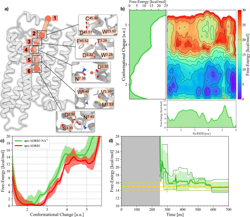
The beta-1 adrenergic receptor (ADRB1) is a critical target for cardiovascular drugs, yet our understanding of how it is activated remains incomplete. Capturing the concerted interplay of agonists, solvent, ions, and protein microswitches is a significant challenge for conventional simulation methods and is essential for unraveling this process. Here, we address this challenge by implementing a powerful enhanced sampling framework that integrates the OneOPES enhanced sampling algorithm with a set of biologically motivated collective variables (CVs). These CVs are designed to track several key features of the activation process simultaneously, including rearrangement of conserved microswitches, the state of the sodium ion binding pocket, and dynamics of critical water molecules. Using this framework, we mapped the multidimensional free energy landscapes of the ADRB1 receptor in both its apo- and adrenaline-bound holo states. Our analysis reveals a detailed, stepwise activation pathway that quantifies the known modulatory roles of sodium ions and protonation states and identifies essential water-mediated networks that stabilize the active conformation. This work provides a detailed overview of ADRB1 activation and establishes the robustness of our OneOPES approach for investigating complex activation mechanisms with the potential for application to other Class A GPCRs.


Figures

1
1
Results of the OneOPES simulations on the apo- and holo-ADRB1 systems. (a) ADRB1 embedded into a POPC/CHL (80:20) model membrane. ADRB1 is colored in gray, whereas POPC and CHL are depicted in orange and cyan, respectively. (b) Binding mode of adrenaline (in yellow) bound to ADRB1. (c) 1D FES as a function of the Conformational Change CV for the apo-ADRB1 systems. (d) Free-energy difference between inactive and active states in the apo-ADRB1 systems over time. In (c,d), the red solid line represents the average of the three replicas, while the transparent red area illustrates the standard deviation. (e) Average 2D FES concerning the RMSD of both the inactive and active structures for the apo-ADRB1 systems. (f) 1D FES as a function of the Conformational Change CV for the holo-ADRB1 systems. (g) Free-energy difference between inactive and active states in the holo-ADRB1 systems over time. In (f,g), the blue solid line represents the average of the three replicas, while the transparent blue area illustrates the standard deviation. (h) Average 2D FES with respect to the RMSD of both the inactive and active structures for the holo-ADRB1 systems. In (d,g), the average value is shown as a yellow dashed line. Error bars of 1 kcal/mol appear as yellow dashed lines. In (e,h), isolines are drawn every 2 kcal/mol.
2
2
Analyses of ADRB1’s microswitches during its apo- and ligand-induced activation. (a) 2D FES as a function of the Conformational Change CV and the respective Distance CVs for the residues of the PIF motif (P5.50–F6.44, I3.40–F6.44, and P5.50–I3.40). (b) 2D FES as a function of the Conformational Change CV for the DRY motif (D3.49–Y34.53), the ionic lock (R3.50–E6.30), and an additional hydrophobic contact in the proximity of the DRY motif (I3.54–L6.34). (c) 2D FES as a function of the Conformational Change CV for the YY motif (Y5.58–Y7.53). On the left side of (a–c), we reported the average 2D FES of the 3 apo-ADRB1 OneOPES simulations, while on the right side, the average 2D FES of the 3 holo-ADRB1 OneOPES simulations. Isolines are drawn every 2 kcal/mol.
3
3
Differences in the internal hydration sites in apo- and holo-ADRB1. (a,b) 2D FES associated with the hydration of (a) apo- and (b) holo-ADRB1’s YY-motif during the GPCR activation. On the right side, insets displaying the water molecules connecting Y5.58 and Y7.53 and the relative frequency of each hydration state in the selected basin. (c) Average distances of N7.45 and N7.53 with respect to the closest water molecule to N7.49 in the apo- and holo-ADRB1 active basins. The image on the right depicts ADRB1 and the phosphorus atoms in the surrounding membrane. The inset displays the water molecule caged between N7.45, N7.49, and Y7.53. d) Distribution of water molecules along ADRB1’s z-axis. Values coming from apo- and holo-ADRB1’s active basins are colored in transparent red and blue, respectively.
4
4
Schematic depiction of the effects of adrenaline binding at ADRB1 and the resulting propagation of structural changes through the receptor. (a) Network of intraprotein contacts that initiates a series of conformational changes extending to the intracellular side upon adrenaline (yellow) binding. (b–f) Average pairwise distances between the pair of residues F6.52–W6.48 (b), N7.45–W6.48 (c), D3.32–Y7.43 (d), N7.45–G7.42 (e), and N7.45–N7.49 (f). Values collected from apo- and holo-ADRB1’s active basins are colored in transparent red and blue, respectively.
5
5
Allosteric modulation of the Na+ binding cavity. (a) Schematic representation of the Na+ translocation pathway (NaPATH CV) within ADRB1. Milestones along the pathway are indicated as orange spheres and shown with inset enlargements for clarity. (b) 2D FES illustrating the relationship between Na PATH (x-axis) and ADRB1’s Conformational Change CV (y-axis). 1D FES projections for the Na PATH and Conformational Change are also shown, providing insights into the energetic landscapes of these processes. (c) 1D FES as a function of the Conformational Change CV for the apo-ADRB1 and apo-ADRB1-NA+ systems. (d) Free-energy difference between inactive and active states in the apo-ADRB1-NA+ system over time. The average value is shown as a yellow dashed line. Error bars of 1 kcal/mol appear as yellow dashed lines.
6
6
Results of the OneOPES simulations on the apo- and holo-ADRB1-ASPH systems. (a,b) 1D FES as a function of the Conformational Change CV for the apo-ADRB1-ASPH (a) and holo-ADRB1-ASPH (b) systems. (c,d) Free-energy difference between inactive and active states in the apo-ADRB1-ASPH (c) and holo-ADRB1-ASPH (d) systems over time. In (a,c), the purple solid line represents the average of the three replicas, while the transparent purple area illustrates the standard deviation. In (b,c), the teal solid line represents the average of the three replicas, while the transparent teal area illustrates the standard deviation. The average value is shown as a yellow dashed line. Error bars of 1 kcal/mol appear as yellow dashed lines. (e,f) Average 2D FES with respect to the RMSD of both the inactive and active structures for the apo-ADRB1-ASPH (e) and holo-ADRB1-ASPH (f) systems.

Similar articles

References

    1. Hilger D., Masureel M., Kobilka B. K.. Structure and dynamics of GPCR signaling complexes. Nat. Struct. Mol. Biol. 2018;25:4–12. doi: 10.1038/s41594-017-0011-7. - DOI - PMC - PubMed
    1. Jong Y.-J. I., Harmon S. K., O’Malley K. L.. GPCR signalling from within the cell. Br. J. Pharmacol. 2018;175:4026–4035. doi: 10.1111/bph.14023. - DOI - PMC - PubMed
    1. Sutkeviciute I., Vilardaga J.-P.. Structural insights into emergent signaling modes of G protein–coupled receptors. J. Biol. Chem. 2020;295:11626–11642. doi: 10.1074/jbc.REV120.009348. - DOI - PMC - PubMed
    1. Lämmermann T., Kastenmüller W.. Concepts of GPCR-controlled navigation in the immune system. Immunol. Rev. 2019;289:205–231. doi: 10.1111/imr.12752. - DOI - PMC - PubMed
    1. Huang Y., Thathiah A.. Regulation of neuronal communication by G protein-coupled receptors. FEBS Lett. 2015;589:1607–1619. doi: 10.1016/j.febslet.2015.05.007. - DOI - PubMed

LinkOut - more resources