Review
. 2025 Jul 28;30(1):94.
doi: 10.1186/s11658-025-00771-1.

Perspectives on mitochondrial dysfunction in the regeneration of aging skeletal muscle

Affiliations
Review

Perspectives on mitochondrial dysfunction in the regeneration of aging skeletal muscle

Kai Wang et al. Cell Mol Biol Lett. .

Abstract

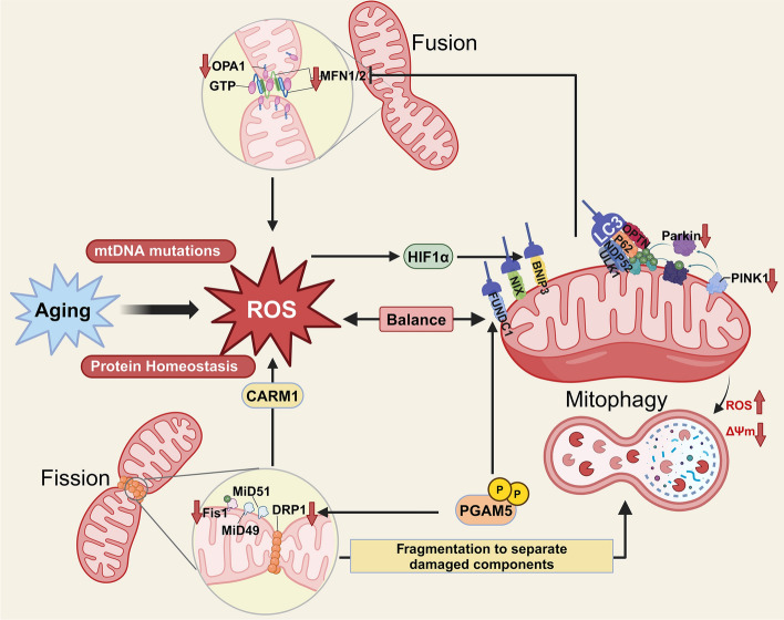
As the global population trends toward aging, the number of individuals suffering from age-related debilitating diseases is increasing. With advancing age, skeletal muscle undergoes progressive oxidative stress infiltration, coupled with detrimental factors such as impaired protein synthesis and mitochondrial DNA (mtDNA) mutations, culminating in mitochondrial dysfunction. Muscle stem cells (MuSCs), essential for skeletal muscle regeneration, also experience functional decline during this process, leading to irreversible damage to muscle integrity in older adults. A critical contributing factor is the loss of mitochondrial metabolism and function in MuSCs within skeletal muscle. The mitochondrial quality control system plays a pivotal role as a modulator, counteracting aging-associated abnormalities in energy metabolism and redox imbalance. Mitochondria meet functional demands through processes such as fission, fusion, and mitophagy. The significance of mitochondrial morphology and dynamics in the mechanisms of muscle regeneration has been consistently emphasized. In this review, we provide a comprehensive summary of recent advances in understanding the mechanisms of aging-related mitochondrial dysfunction and its role in hindering skeletal muscle regeneration. Additionally, we present novel insights into therapeutic approaches for treating aging-related myopathies.

Keywords: Aging; Mitochondrial dynamics; Mitophagy; Oxidative stress; Skeletal muscle regeneration.


Conflict of interest statement

Declarations. Ethical approval and consent to participate: Not applicable. Consent for publication: Not applicable. Competing interests: The authors declare no potential conflicts of interest.

Figures

Fig. 1
Fig. 1
Mitochondrial dynamics in muscle regeneration: insights from young and aged individuals. The figure was created using Biorender (https://www.biorender.com/). In young individuals, mitochondria facilitate muscle regeneration by exhibiting efficient energy responses, thereby promoting the proliferation and activation of MuSCs. This process induces temporally and spatially specific gene expression patterns, with multiple epigenetic regulatory factors contributing to its modulation. By contrast, aging impairs MuSC regenerative capacity by disrupting mitochondrial homeostasis and various metabolic processes. Excessive accumulation of ROS and the release of mtDNA exacerbate cellular damage, while progressive energy deficits hinder MuSC self-renewal and activation
Fig. 2
Fig. 2
Aging-mediated disorders of skeletal muscle regeneration. The figure was created using Biorender (https://www.biorender.com/). Aging disrupts proteostasis and promotes unfavorable posttranslational modifications, leading to the accumulation of ROS. This, in turn, forms a detrimental feedback loop with mitochondrial dysfunction, collectively impairing the regenerative capacity of aged skeletal muscle. Moreover, aging alters cellular vitality and epigenetic landscapes in response to various signaling factors. It also remodels the stem cell niche, suppressing the activation potential ofMuSCs
Fig. 3
Fig. 3
Mitochondrial functions and postdamage effects of the aging process. The figure was created using Biorender (https://www.biorender.com/). Sirtuin (SIRT)2, SIRT3, and SIRT7 are several members of the Sirtuin family of NAD+-dependent deacetylases that play a role in stem cell mitochondrial protection, and the expression of these sirtuins is inhibited in stem cells during senescence. The absence of these sirtuins leads to mitochondrial stress and functional decline, and this loss is a mitochondrial stress and senescence driver of stem cell functional degradation and a control node for rejuvenating senescent stem cells [–123]. The effects over time are enormous. α-klotho is an anti-aging protein, but the loss of its promoter demethylation modification with age plays an important role in stem cell dysfunction, as demonstrated in the study of MuSCs, where the loss of its content has an inhibitory effect on the assembly of the mitochondrial respiratory chain and on mitochondrial function, leading to a shift in the mechanisms of skeletal muscle regeneration [124]. Mitochondrial dysfunction in aging, characterized by mtDNA damage, directly contributes to the development of muscle regenerative dysfunction [125] and changes in the metabolic response to mitochondrial complex I in myoblast myogenic programmed differentiation, and supplementation with NAD to improve mitochondrial function could be a possible option in ameliorating the impaired function of MuSCs caused by aging or disease [126]. Alterations in mitochondria and metabolic capacity by sepsis, a typical toxic effect leading to muscle weakness, underlie the induction of long-term damage to satellite cells and lead to inefficient muscle regeneration and thus muscle strength [127]. Mitochondrial dysfunction in MuSCs often leads to impaired muscle regeneration in the aggregate, implying that in the state of impaired regenerative function of muscles due to aging mitochondria and MuSCs senescence may have some kind of interactive relationship, and mitochondria may act as a bridge to help the damage of senescence on MuSCs
Fig. 4
Fig. 4
Mitochondria drive metabolic reprogramming in skeletal muscle regeneration. The figure was created using Biorender (https://www.biorender.com/). Quiescent satellite cells (QSCs) preferentially utilize fatty acids to meet their low energy demands. The metabolic intermediate acetyl-CoA supports histone H4K16 acetylation, which is crucial for maintaining MuSC quiescence and self-renewal capacity. In contrast, ASCs rely on glucose metabolism to rapidly meet the increased energy requirements of MuSCs. Through OxPhos, ASCs generate higher levels of ATP and ROS to support proliferation and differentiation. Furthermore, proliferating muscle satellite cells (MuSCs) exhibit heightened histone acetylation levels, mechanistically linked to their metabolic reprogramming toward increased lactate-producing glycolytic flux and concomitant reduction in SIRT1 deacetylase activity. This dual regulation elevates intracellular acetyl-CoA pools, creating an epigenetic permissive state that licenses transcriptional activation of proproliferative gene programs
Fig. 5
Fig. 5
Crosstalk between oxidative stress and mitochondrial quality control in aged MuSCs. The figure was created using Biorender (https://www.biorender.com/). Mitochondria are the primary sites of ROS production. Aging contributes to ROS accumulation through mtDNA mutations and disrupted proteostasis. In response, the mitochondrial quality control (MQC) system actively mitigates ROS levels by modulating mitochondrial fission and employing mitophagy to clear damaged mitochondria. However, excessive ROS overwhelms the MQC system, leading to its dysfunction. Age-associated deficiencies in key regulators such as optic nerve atrophy protein 1 (OPA1) and dynamin-related protein (DRP1) further exacerbate mitochondrial quality defects, amplifying ROS-induced cellular damage and impairing mitochondrial homeostasis

References

    1. Dumont NA, Wang YX, Rudnicki MA. Intrinsic and extrinsic mechanisms regulating satellite cell function. Development. 2015;142(9):1572–81. 10.1242/dev.114223. - PMC - PubMed
    1. Seale P, Asakura A, Rudnicki MA. The potential of muscle stem cells. Dev Cell. 2001;1(3):333–42. 10.1016/s1534-5807(01)00049-1. - PubMed
    1. Mauro A. Satellite cell of skeletal muscle fibers. J Biophys Biochem Cytol. 1961;9(2):493–5. 10.1083/jcb.9.2.493. - PMC - PubMed
    1. Scharner J, Zammit PS. The muscle satellite cell at 50: the formative years. Skelet Muscle. 2011;1:28. 10.1186/2044-5040-1-28. - PMC - PubMed
    1. Short KR, Bigelow ML, Kahl J, Singh R, Coenen-Schimke J, Raghavakaimal S, et al. Decline in skeletal muscle mitochondrial function with aging in humans. Proc Natl Acad Sci USA. 2005;102(15):5618–23. 10.1073/pnas.0501559102. - PMC - PubMed