. 2025 Jul 28;23(1):849.
doi: 10.1186/s12967-025-06827-2.

Novel loop structure of human IgG1 Fc fused CD38 targeted bispecific antibodies and their anti-tumor effect in acute myeloid leukemia

Affiliations

Novel loop structure of human IgG1 Fc fused CD38 targeted bispecific antibodies and their anti-tumor effect in acute myeloid leukemia

Shisen Wang et al. J Transl Med. .

Abstract

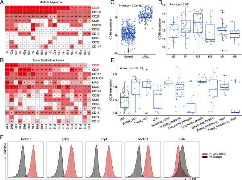
Background: Acute myeloid leukemia (AML) still lacks an ideal immunotherapy target. CD38 serves as a potential therapeutic target for AML. Classical bispecific antibody (BsAb) requires continuous infusion due to small molecular size and short half-life.

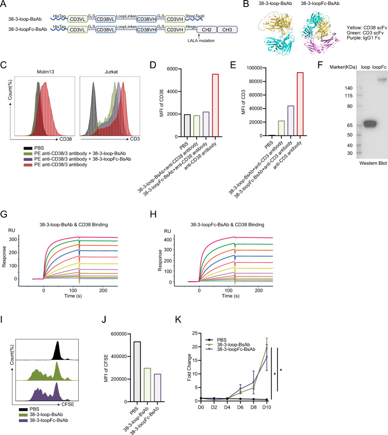
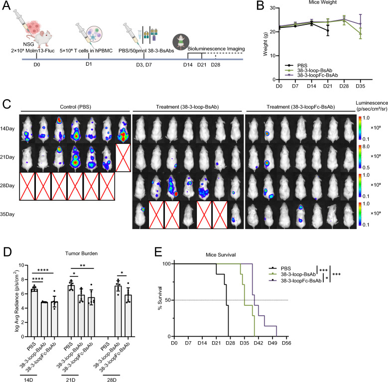
Methods: The anti-human CD38 single-chain variable fragment (scFv) and anti-human CD3 scFv were cloned to expression plasmids. ExpiCHO-S cells were transfected, and the anti-CD38/anti-CD3 bispecific antibody (38-3-BsAbs) in CHO supernatants was efficiently purified. The anti-AML efficacy of 38-3-BsAbs was evaluated in vitro and in vivo.

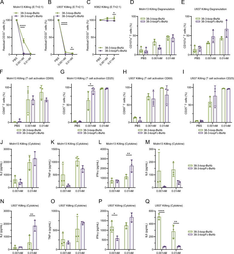
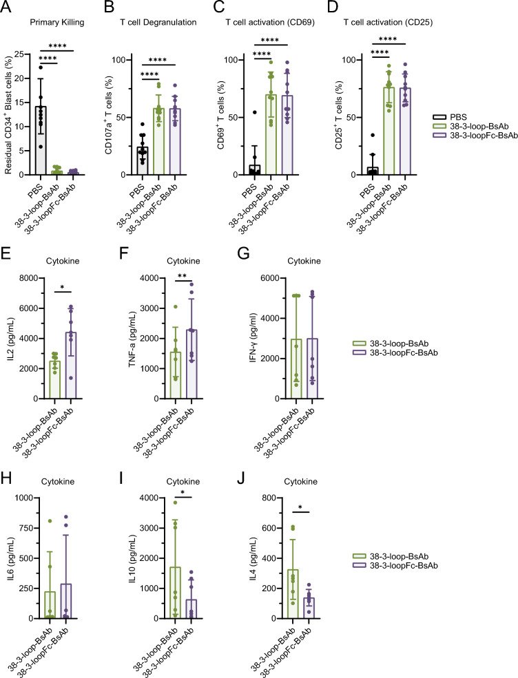
Results: The novel loop structures of non-human IgG Fc fused and human IgG1 Fc fused anti-CD38/anti-CD3 BsAbs, 38-3-loop-BsAb and 38-3-loopFc-BsAb, were designed and produced. Both 38-3-loop-BsAb and 38-3-loopFc-BsAb showed excellent anti-AML effects at low concentrations in vitro. AML xenograft NOD-scid IL2gammanull (NSG) mouse model was adopted to evaluate therapeutic effects of 38-3-BsAb. The anti-leukemic effect of 38-3-loopFc-BsAb was superior.

Conclusions: We report on new structures of 38-3 bispecific antibody and demonstrate their anti-tumor effect in the treatment of AML.

Keywords: Acute myeloid leukemia; Bispecific antibody; CD38; Immunotherapy.


Conflict of interest statement

Declarations. Ethics approval and consent to participate: This study of patients and animal experiments was approved by the Ethics Review Board of IHCAMS. All patients and health donors were informed, and written consents were signed in accordance with the Declaration of Helsinki. Consent for publication: Not applicable. Competing interests: The authors declare that they have no competing interests.

Figures

Fig. 1
Fig. 1
CD38 is a potential immunotherapeutic target of acute myeloid leukemia. A Heatmap of common flow cytometry markers of 20 multiple myeloma (MM) patients in IHCAMS. B Heatmap of common flow cytometry markers of 20 acute myeloid leukemia (AML) patients in IHCAMS. +  +  + strongly positive, +  + positive, + dim, −negative, grey represents data not available. C Box plot comparing mRNA expression level of CD38 between normal people and LAML patients. Normal data from GTEx database and TCGA-LAML expression data are merged. Batch effect is removed. D Box plot showing mRNA expression level of CD38 in TCGA-LAML dataset based on French American British (FAB) classification. E Box plot showing mRNA expression level of CD38 in different hematologic malignancy cell lines in CCLE database. F Flow cytometry histograms showing CD38 expression intensity among different AML cell lines, red represents CD38 intensity, black represents isotype control
Fig. 2
Fig. 2
Construction, production, and biological functions of 38-3 bispecific antibodies. A Schematic diagram of 38-3-loop-BsAb and 38-3-loopFc-BsAb. B 38-3-BsAb protein structures predicted with AlphaFold2. Yellow represents CD38 scFv. Green represents CD3 scFv. Purple represents mutated IgG1 Fc. C Flow cytometry histograms showing competitive combination of 38-3-BsAbs with commercial flow cytometry antibodies on CD38+ Molm13 and CD3+ Jurkat. Left, PE anti-CD38 antibody. Right, PE anti-CD3 antibody. D, E Box plot showing Mean Fluorescence Intensity (MFI) of competitive combination in Figure C. The MFI of CD38 from left to right was 1970, 1878, 2204, and 5544, respectively. The MFI of CD3 from left to right was 833, 22010, 44146, and 93271, respectively. F Western blot showing the actual molecular size of 38–3-BsAbs. G, H SPR binding assay demonstrated efficient binding of 38-3-loop-BsAb (G) and 38–3-loopFc-BsAb (H) with CD38. BsAbs were diluted into 12 gradients from the highest concentration (38-3-loop-BsAb: 360nM; 38-3-loopFc-BsAb: 500nM). 38-3-loop-BsAb & CD38: 1:1 Binding, ka = 3.27e5, kd = 5.88e−5, Rmax = 285.9, KD = 1.80e−10; 38-3-loopFc-BsAb & CD38: 1:1 Binding, ka = 1.68e5, kd = 6.12e−5, Rmax = 368.4, KD = 3.64e−10. I Flow cytometry histogram showing CFSE dye fluorescence attenuation of primary T cell after stimulated by 38–3-BsAbs for 5 days. J Box plot showing Mean Fluorescence Intensity (MFI) of CFSE dye fluorescence attenuation in Figure I. The MFI of CFSE from left to right was 531529, 297970, and 245529, respectively. K Line graph illustrating the fold change of the primary T cells’ proliferation after stimulation by the 38-3-BsAbs
Fig. 3
Fig. 3
38-3-BsAbs induce potent lysis of CD38+ AML cell lines by primary T cell from healthy donors. AC Specific lysis of CD38+ and CD38 target cells mediated by primary T cells (E:T ratio of 2:1) after 72 h of co-culture assay at different 38-3-BsAb concentrations. D, E Box plot showing the degranulation (CD107a) of T cells in the specific killing mediated by 38-3-BsAb. FI Box plot showing the activation (CD69, CD25) of T cells in the killing assay of Molm13 and U937. (J-Q) Box plots comparing cytokine release (IL2, TNF-a, IFN-γ, IL6) when primary T cells kill CD38+ target cells. The cytokine release level of PBS group was very low, which was not shown
Fig. 4
Fig. 4
38-3-BsAbs mediate effective lysis of primary CD38+ AML blast cells. A Specific lysis of primary CD38+ AML blast cells cocultured with health donor T cells (E:T ratio of 2:1) for 72 h in the presence of 0.1nM 38-3-BsAbs. BD Box plot showing the degranulation (CD107a) and activation (CD69, CD25) of T cells in the killing assay. EJ Box plots showing the cytokine release (IL2, TNF-a, IFN-γ, IL6, IL10, and IL4) in the killing assay. The cytokine release level of PBS group was very low, which was not shown
Fig. 5
Fig. 5
38-3-BsAbs exert good anti-AML efficacy in vivo. A Schematic diagram of in vivo assay. B Line graph showing the change of the mice weight. C Bioluminescence imaging of mice in control group and 38-3-BsAbs treatment groups since week 2. D Box plot comparing mice tumor burden among three groups on week 2, 3, 4, 5. Log Average radiance quantification (p/sec/cm2) of bioluminescence imaging was used as a relative reflection of tumor burden. E Kaplan–Meier plot showing the survival of mice in three groups
Fig. 6
Fig. 6
Impact of 38-3-BsAbs on T cells engraftment and persistence, and systemic cytokine levels monitoring in mice. A Schematic diagram of the continuous monitoring. B Line graph showing the changes of the T-cell proportion in mice PBMC (mPBMC) after PBS or 38-3-BsAbs dosing. Statistical significance of key time points (day1, 7, 17) is annotated. C Box plot comparing T-cell proportion in mice BMMC (mBMMC) among the PBS or 38-3-BsAbs treatment group on day 7 and day 15. D, E Line graph showing the cytokine monitoring of IFN-γ and IL10. Statistical significance of key time points (IFN-γ: day1, 3, 9, 13; IL10: day1) is annotated. F Line graph showing dynamics of 38-3-BsAbs concentrations in vivo after dosing

Similar articles

References

    1. Döhner H, Weisdorf Daniel J, Bloomfield Clara D. Acute myeloid leukemia. N Engl J Med. 2015;373:1136–52. - PubMed
    1. Kantarjian HM, DiNardo CD, Kadia TM, Daver NG, Altman JK, Stein EM, Jabbour E, Schiffer CA, Lang A, Ravandi F. Acute myeloid leukemia management and research in 2025. CA Cancer J Clin. 2025;75:46–67. - PMC - PubMed
    1. Jabbour E, Short NJ, Jain N, Haddad FG, Welch MA, Ravandi F, Kantarjian H. The evolution of acute lymphoblastic leukemia research and therapy at MD Anderson over four decades. J Hematol Oncol. 2023;16:22. - PMC - PubMed
    1. Restelli C, Ruella M, Paruzzo L, Tarella C, Pelicci PG, Colombo E. Recent advances in immune-based therapies for acute myeloid leukemia. Blood Cancer Discov. 2024;5:234–48. - PMC - PubMed
    1. van de Donk NWCJ, Pawlyn C, Yong KL. Multiple myeloma. Lancet (London, England). 2021;397:410–27. - PubMed

MeSH terms