

Licochalcone D inhibits osteoclast differentiation and postmenopausal osteoporosis by inactivating the NF-κB signaling pathway
- PMID: 40722101
- PMCID: PMC12302740
- DOI: 10.1186/s13018-025-06132-0
Licochalcone D inhibits osteoclast differentiation and postmenopausal osteoporosis by inactivating the NF-κB signaling pathway
Abstract
Background: Osteoporosis is prevalent among postmenopausal women and is characterized by excessive bone resorption primarily mediated by osteoclasts. This study aimed to investigate the effects of the natural compound Licochalcone D (Lico D) on osteoclast differentiation and its therapeutic potential in ovariectomized (OVX) mouse models of osteoporosis.
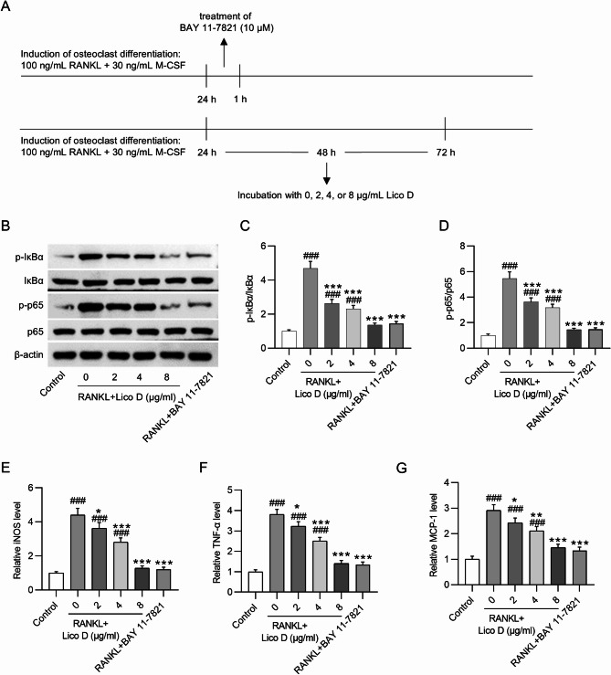
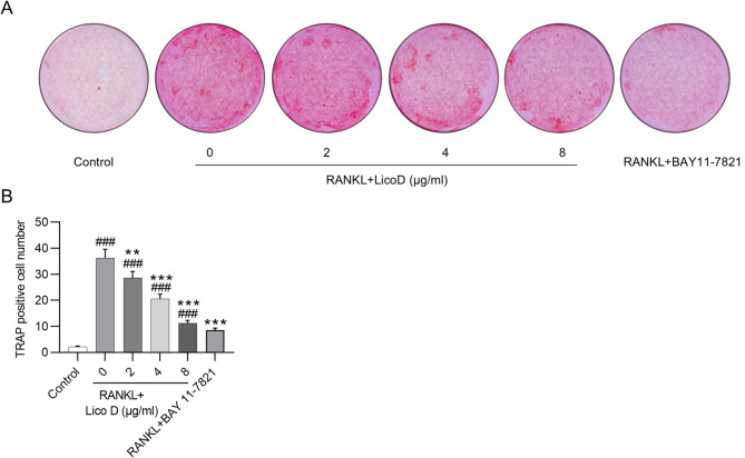
Methods: The cytotoxicity of various doses of Lico D on mouse bone marrow-derived macrophages (BMMs) was evaluated using CCK-8 assays. The differentiation of BMMs into osteoclasts was induced by RANKL treatment, followed by exposure to Lico D at doses of 2, 4, and 8 µg/ml. Additionally, 10 µM BAY 11-7821 (an NF-κB inhibitor) was used to inhibit NF-κB signaling in RANKL-stimulated BMMs. TRAP staining was conducted to measure osteoblast cell number. Western blot analysis was performed to measure protein levels of osteoclast differentiation markers and NF-κB-related factors. RT-qPCR was performed to assess the mRNA levels of downstream genes in the NF-κB pathway. In animal experiments, OVX mice received intraperitoneal injections of Lico D at doses of 10 or 50 mg/kg. Subsequently, femurs were harvested for histopathological examination.
Results: Lico D at doses of 2-8 µg/ml showed no significant cytotoxicity toward BMMs. In addition, Lico D inhibited RANKL-induced osteoclast formation and downregulated protein levels of osteoclast-specific genes (mmp9, ctsk, c-Fos and nfatc1). Moreover, Lico D suppressed the phosphorylation of NF-κB p65 and IκBα in RANKL-treated BMMs. Importantly, the suppressive effects of Lico D, especially at 8 µg/ml, on osteoclast cell number and osteoclast-specific markers were comparable to BAY 11-7821. Moreover, Lico D inhibited OVX-induced bone loss and restored dysregulated bone parameters in mice.
Conclusion: Lico D inhibits RANKL-induced osteoclast differentiation and alleviates postmenopausal osteoporosis in mice by suppressing the NF-κB signaling pathway.
Keywords: BAY 11-7821; Bone marrow-derived macrophages; Licochalcone D; NF-κB signaling; Osteoporosis.
© 2025. The Author(s).
Conflict of interest statement
Declarations. Ethical approval: The ethics committee of The Third Affiliated Hospital of Soochow University approved all animal experiments. Competing interests: The authors declare no competing interests.
Figures


Similar articles
-
Yougui pills prevent ovariectomy-induced bone loss by suppressing Th17 response and IL-17/NF-κB pathway.Ann Med. 2025 Dec;57(1):2529576. doi: 10.1080/07853890.2025.2529576. Epub 2025 Jul 7. Ann Med. 2025. PMID: 40622369 Free PMC article.
-
Anti-osteoporosis effect of Semen Cuscutae in ovariectomized mice through inhibition of bone resorption by osteoclasts.J Ethnopharmacol. 2022 Mar 1;285:114834. doi: 10.1016/j.jep.2021.114834. Epub 2021 Nov 18. J Ethnopharmacol. 2022. PMID: 34801609
-
Tectorigenin Ameliorates Glucocorticoid-Induced Osteoporosis by Inhibiting the NF-κB Signal Pathway and Modulating Treg-Th17 Cell Balance.J Cell Mol Med. 2025 Jul;29(13):e70705. doi: 10.1111/jcmm.70705. J Cell Mol Med. 2025. PMID: 40619720 Free PMC article.
-
Docosahexaenoic Acid Inhibits Osteoclastogenesis via FFAR4-Mediated Regulation of Inflammatory Cytokines.Molecules. 2025 Jul 29;30(15):3180. doi: 10.3390/molecules30153180. Molecules. 2025. PMID: 40807354 Free PMC article. Review.
-
Oxidative stress and inflammation: roles in osteoporosis.Front Immunol. 2025 Aug 12;16:1611932. doi: 10.3389/fimmu.2025.1611932. eCollection 2025. Front Immunol. 2025. PMID: 40873591 Free PMC article. Review.
References
-
- Conti V, Russomanno G, Corbi G, Toro G, Simeon V, Filippelli W, Ferrara N, Grimaldi M, D’Argenio V, Maffulli N, Filippelli A. A polymorphism at the translation start site of the vitamin D receptor gene is associated with the response to anti-osteoporotic therapy in postmenopausal women from Southern Italy. Int J Mol Sci. 2015;16:5452–66. - PMC - PubMed
MeSH terms
Substances
Grants and funding
LinkOut - more resources
Full Text Sources
Miscellaneous
