

Analysing plate fixation of a comminuted fracture of the proximal ulna in relation to the elbow joint: a finite element study
- PMID: 40722181
- PMCID: PMC12302834
- DOI: 10.1186/s13018-025-06031-4
Analysing plate fixation of a comminuted fracture of the proximal ulna in relation to the elbow joint: a finite element study
Abstract
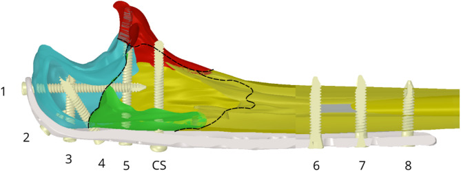
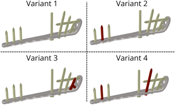
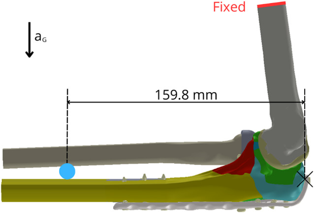
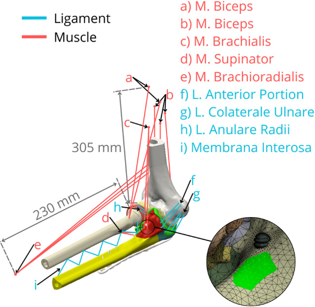
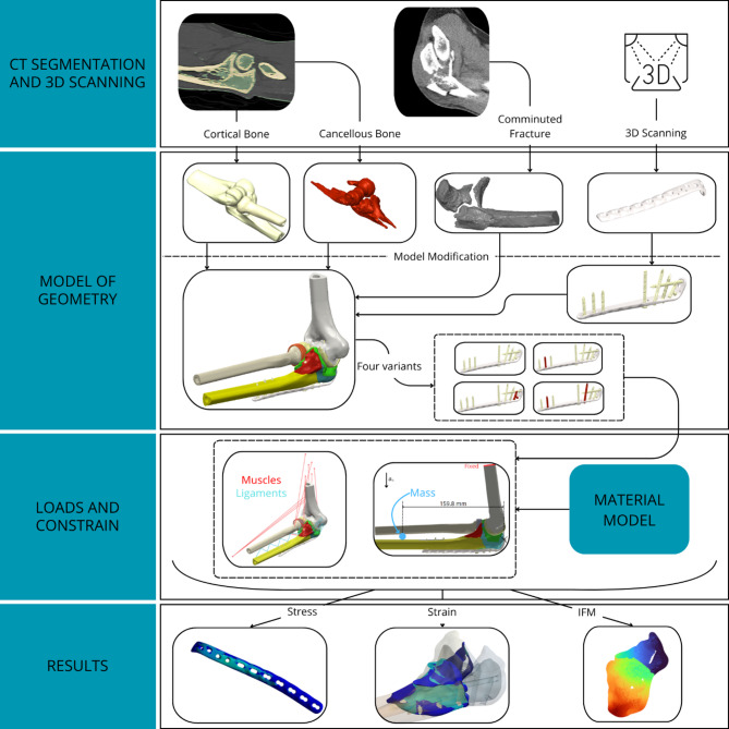
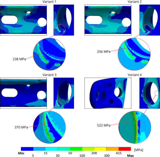
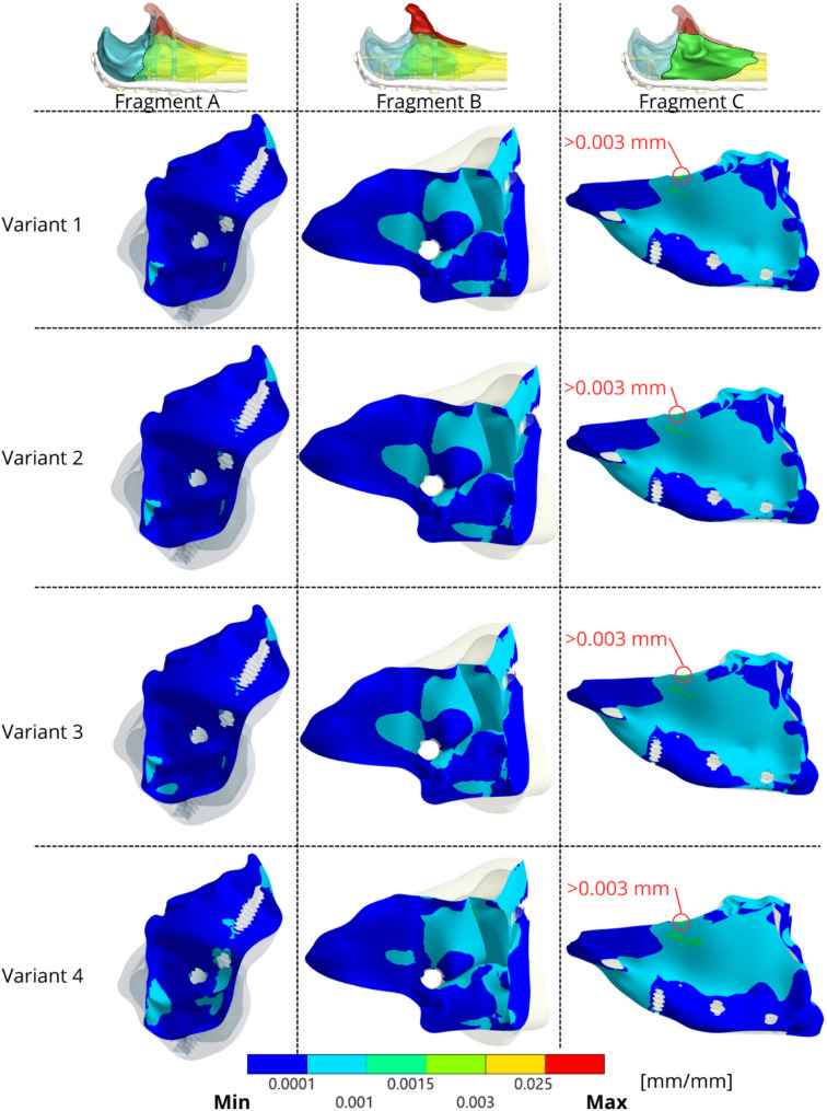
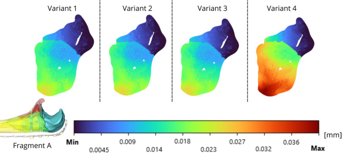
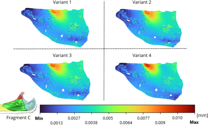
This study investigated the biomechanical behavior of four different screw configurations used to fix comminuted proximal ulna fractures with a locking compression plate (LCP), via a detailed finite element model based on realistic anatomical geometry. The model incorporated realistic anatomical geometry including both cortical and cancellous bone, soft tissue constraints, and loading conditions representing the physiological self-weight of the forearm, with the humerus fixed at its proximal end. The stress distribution on the plate, strain intensity within the bone tissue, and interfragmentary motion (IFM) between fracture fragments were evaluated for each configuration. The results indicate that all the tested configurations provide adequate stability under normal loading conditions, with no risk of material failure. However, excessive stress concentrations were observed in specific screw regions depending on the configuration, particularly when proximal screws anchoring the olecranon (e.g. screws 2 and 3 in Variant 3) were omitted. Strain analysis revealed moderate physiological bone loading across variants, whereas IFM assessment highlighted the importance of securing the coronoid and apical fragments to prevent compromised healing. These findings suggest that a specific reductions in osteosynthetic material, such as omitting certain diaphyseal screws while maintaining crucial olecranon and coronoid fixation, may provide sufficient fracture stabilisation under the modelled conditions, potentially minimising implant-related complications. This modelling approach offers a valuable tool for preclinical assessment of osteosynthesis strategies and supports future comparative research on fixation methods with varying biomechanical properties.
Keywords: Elbow joint; Finite element analysis; Interfragmentary motion; Locking compression plate; Olecranon fracture; Proximal ulna comminuted fracture.
Conflict of interest statement
Declarations. Competing interests: The authors declare no competing interests.
Figures


Similar articles
-
Surgical interventions for treating fractures of the olecranon in adults.Cochrane Database Syst Rev. 2014 Nov 26;2014(11):CD010144. doi: 10.1002/14651858.CD010144.pub2. Cochrane Database Syst Rev. 2014. PMID: 25426876 Free PMC article.
-
High Failure Rates in Comminuted Patella Fractures (AO/OTA 34-C3) Fixed With an Isolated, New Patella-Specific 2.7-mm Variable-Angle Locking Plate.J Orthop Trauma. 2025 Jun 1;39(6):320-330. doi: 10.1097/BOT.0000000000002972. J Orthop Trauma. 2025. PMID: 40052806
-
Efficacy evaluation of Kirschner wire tension band combined with anatomical locking plate in the treatment of Mayo type II olecranon fractures.BMC Musculoskelet Disord. 2025 Jul 4;26(1):597. doi: 10.1186/s12891-025-08843-1. BMC Musculoskelet Disord. 2025. PMID: 40616018 Free PMC article. Clinical Trial.
-
Is Tension Band Wire Fixation Superior to Plate Fixation for Simple Displaced Olecranon Fractures? A Randomized Trial With Median Follow-up of 7.5 Years.Clin Orthop Relat Res. 2024 Jan 1;482(1):127-133. doi: 10.1097/CORR.0000000000002832. Epub 2023 Sep 7. Clin Orthop Relat Res. 2024. PMID: 37678389 Free PMC article. Clinical Trial.
-
Trans-ulnar fracture dislocations of the elbow: a systematic review and clarification of classification systems.J Shoulder Elbow Surg. 2024 Apr;33(4):975-983. doi: 10.1016/j.jse.2023.10.014. Epub 2023 Nov 29. J Shoulder Elbow Surg. 2024. PMID: 38036255
References
-
- Court-Brown CM, Caesar B. Epidemiology of adult fractures: A review. Injury. Aug. 2006;37(8)691–697 10.1016/j.injury.2006.04.130 - PubMed
-
- Duckworth AD, Clement ND, Aitken SA, Court-Brown CM, McQueen MM. The epidemiology of fractures of the proximal ulna. Injury. Mar. 2012;43(3):343–346. 10.1016/j.injury.2011.10.017 - PubMed
-
- Wilkerson JA, Rosenwasser MP. Surgical techniques of olecranon fractures. J Hand Surg Am. Aug. 2014;39(8):1606–14. 10.1016/j.jhsa.2014.05.014. - PubMed
-
- Tarallo L, Mugnai R, Adani R, Capra F, Zambianchi F, Catani F. Simple and comminuted displaced olecranon fractures: A clinical comparison between tension band wiring and plate fixation techniques, Arch Orthop Trauma Sur. Aug. 2014;134(8):1107–1114. 10.1007/s00402-014-2021-9 - PubMed
Grants and funding
LinkOut - more resources
Full Text Sources
