Review
. 2025 Jul 3;15(7):716.
doi: 10.3390/brainsci15070716.

Looking into Abnormal Co-Expressions of Tau and TDP-43 in the Realm of Mixed Dementia Types: A Double-Punch Scenario

Affiliations
Review

Looking into Abnormal Co-Expressions of Tau and TDP-43 in the Realm of Mixed Dementia Types: A Double-Punch Scenario

Hossam Youssef et al. Brain Sci. .

Abstract

Transactive response DNA-binding protein of 43 kDa (TDP-43) and tau proteins play critical roles in neurodegenerative diseases, particularly frontotemporal lobar degeneration (FTLD) and Alzheimer's disease (AD). The co-occurrence of TDP-43 and tau pathologies raises questions about their role in disease progression. This review explores the simultaneous presence of tau and TDP-43 co-pathologies, emphasizing their molecular interactions and the resultant neuropathological implications. Additionally, we provide representative examples of their clinical presentations, neuroimaging, and neuropathological findings associated with FTLD-TDP and FTLD-tau, emphasizing the need for a comprehensive understanding of these intertwined pathologies. We analyze various clinical scenarios, including argyrophilic grain disease (AGD), primary age-related tauopathy (PART), and limbic predominant age-related TDP-43 encephalopathy (LATE), to elucidate the complex relationship between these proteinopathies. From the literature, the co-occurrence of tau and TDP-43 is linked to more severe and poorer clinical outcomes compared to isolated pathologies. This review underscores the necessity of considering co-pathologies in the context of FTLD, as they may act as accelerators of cognitive decline. This highlights the importance of integrated approaches in diagnosing and treating neurodegenerative conditions characterized by tau and TDP-43 misfolding. Understanding the interplay between these molecular markers is vital for advancing therapeutic strategies for such disorders.

Keywords: Alzheimer’s disease; TDP-43; biomarkers; frontotemporal lobar degeneration; molecular mechanisms; neuroimaging; neuropathology; tau.


Conflict of interest statement

The authors declare no conflicts of interest.

Figures

Figure 1
Figure 1
Schematic representation of a neuron under normal and pathological conditions as described in mixed proteinopathies. Key differences between physiological (A) and pathological (B) conditions include the altered localization of TDP-43, its contribution to tau mRNA instability and the 3R/4R tau ratio, as well as the post-translational modifications observed in tau and TPD-43 in the pathological state including phosphorylations, among others. Note synergistic interactions that drive the misfolding of both proteins.
Figure 2
Figure 2
The diagram shows the relationship between the main clinical and pathological attributes in the dementia spectrum and their underlying biochemical features. The relationship between chemical biomarkers (pink boxes) and clinical (orange boxes) or neuropathological (green boxes) phenotypes is pointed out by orange arrows. The possible mixed clinicopathological presentations are marked by the yellow arrows. Note the co-expression of tau and TDP-43 can be observed in the context of Alzheimer’s disease (AD). as well as in Frontotemporal lobar degeneration (FTLD). These mixed types of dementias tend to commonly occur in the elderly where an increased incidence of TDP aggregates in situations like limbic-predominant age-related TDP-43 encephalopathy (LATE) or increasing tau aggregates in aging-related tau astrogliopathy (ARTAG), Primary Age-Related Tauopathy (PART) are present. Abbreviations: pTAU, misfolded phosphorylated tau protein; β-Amyl, misfolded beta-amyloid protein; pTDP-43, misfolded phosphorylated TDP-43 protein; 4-TAU, abnormal tau protein isoform with four microtubule-binding domains; 3-TAU, abnormal tau protein isoform with three microtubule-binding domains; FTLD-TDP, Frontotemporal lobar degeneration with TDP-43 pathology; FTLD-tau, Frontotemporal lobar degeneration with tau pathology; bvFTD, behavioral variant of frontotemporal dementia; PPA, primary progressive aphasia; PSP, progressive supranuclear palsy; CBD, cortico-basal degeneration; AGD, argyrophilic grain disease; PiD, Pick’s disease.
Figure 3
Figure 3
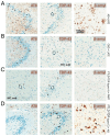
Representative neuropathological examples of mixed tau and TDP proteinopathies. (A) Immunohistochemical staining of the hippocampal dental gyrus staining with phosphorylated tau (AT8) and phosphorylated TDP (TDP-43), which are shown in white arrows. Additional beta-amyloid (β-amyl) staining from the inferior temporal cortex is presented. Other co-pathologies include CBD + AD (B), FTLD-TDP-43 (type a) +PART (C), and FTLD-TDP-43(type c) +AD (D). Hematoxylin is used as a nuclear counterstaining (blue). Scale bar = 100 microns.
Figure 4
Figure 4
Clinical neuroimaging examples of microstructural white matter changes in a subgroup of illustrative cases with multi-dementia expressing abnormal tau and TDP-43. (A) Diffusion tensor imaging-based tractography representations on different views along the left uncinate fasciculi (in yellow). (B) Fractional anisotropy (FA) from these tracts where decreased (increased microstructural worsening) and worse in cases expressing simultaneously phosphorylated tau and TDP-43. (C) Gallyas-silver staining across representative white matter in the left temporal regions from subjects with premortem neuroimaging displayed in (B), showing increased microstructural alterations in neurofilament organization in patients with tau and TDP-43 multi-pathologies. Note that the comparison was conducted based on subjects with equal ADNC scores and comparable premortem imaging, as well as the length of the disease.
Figure 5
Figure 5
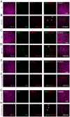
Immunohistochemical patterns of temporal white matter regions by fluorescence confocal microscopy from a short representative set of patients diagnosed with mixed dementias. (A) Confocal evaluation of temporal white matter (WM) tracts by neurofilament heavy chain (NF-H) markers (magenta), phosphorylated tau (AT8 in green), and phosphorylated TDP-43 (red) is shown from a patient with AD. Note a decrease in neurofilament content in patients with AD and LATE (B). A sample from a subject with CBD (C) showed a more preserved WM microstructure than a patient with CBD and LATE (D). Similar increasing alterations were seen between FTLD-TDP subjects with the additional presence of PART (E vs. F), as well as AD (G vs. H) co-pathologies. Note the increased co-localization between AT8 and TDP-43 (asterisk), which could indicate a coexistent burden mechanism. Scale bar = 20 microns.
Figure 6
Figure 6
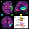
Selected clinical examples to illustrate molecular imaging changes of mixed dementia pathologies by tau-PET imaging. (A) Representative axial, lateral, and coronal views by tau-PET imaging and automatic segmentation from regions of interest (ROIs) including the superior (blue), middle (green), and inferior (yellow) left temporal lobe. (B) Quantitative evaluation of the average tau-PET bindings from the combined left temporal ROIs by standardized uptake ratio (SUVR). Considering comparable ADNC scores between subjects with single and dual pathology, we observed a larger amount of tau-deposition in subjects with co-pathologies. This may indicate that the presence of TDP-43 could enhance the deposition by tau-PET bindings in such complex cases.

Similar articles

References

    1. Gefen T., Ahmadian S.S., Mao Q., Kim G., Seckin M., Bonakdarpour B., Ramos E.M., Coppola G., Rademakers R., Rogalski E., et al. Combined Pathologies in FTLD-TDP Types A and C. J. Neuropathol. Exp. Neurol. 2018;77:405–412. doi: 10.1093/jnen/nly018. - DOI - PMC - PubMed
    1. Latimer C.S., Liachko N.F. Tau and TDP-43 synergy: A novel therapeutic target for sporadic late-onset Alzheimer’s disease. Geroscience. 2021;43:1627–1634. doi: 10.1007/s11357-021-00407-0. - DOI - PMC - PubMed
    1. Meneses A., Koga S., O’Leary J., Dickson D.W., Bu G., Zhao N. TDP-43 Pathology in Alzheimer’s Disease. Mol. Neurodegener. 2021;16:84. doi: 10.1186/s13024-021-00503-x. - DOI - PMC - PubMed
    1. Nelson P.T., Fardo D.W., Wu X., Aung K.Z., Cykowski M.D., Katsumata Y. Limbic-predominant age-related TDP-43 encephalopathy (LATE-NC): Co-pathologies and genetic risk factors provide clues about pathogenesis. J. Neuropathol. Exp. Neurol. 2024;83:396–415. doi: 10.1093/jnen/nlae032. - DOI - PMC - PubMed
    1. Pennington C., Marini L., Coulthard E., Love S. Mixed neuropathology in frontotemporal lobar degeneration. Amyotroph. Lateral Scler. Front. Degener. 2020;21:301–308. doi: 10.1080/21678421.2020.1733019. - DOI - PubMed

LinkOut - more resources