

Performance of Computed Tomography of the Kidneys, Ureter and Bladder in Non-Calculus Diagnoses: A Comparative Review of Non-Enhanced with Intravenous Contrast-Enhanced Imaging
- PMID: 40722481
- PMCID: PMC12293321
- DOI: 10.3390/diagnostics15141731
Performance of Computed Tomography of the Kidneys, Ureter and Bladder in Non-Calculus Diagnoses: A Comparative Review of Non-Enhanced with Intravenous Contrast-Enhanced Imaging
Abstract
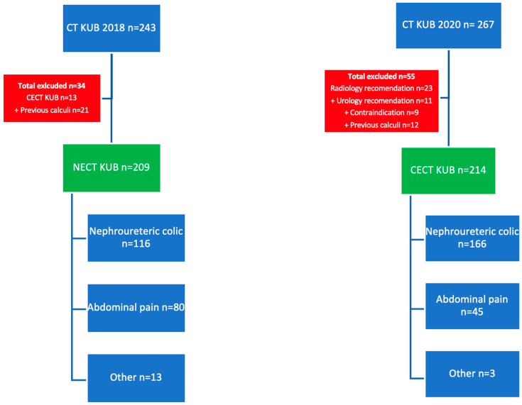
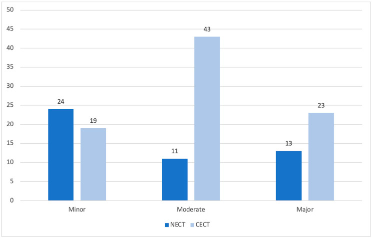
Background/Objectives: Non-enhanced computed tomography of the kidneys, ureters and bladder (NECT KUB) is the initial imaging modality for suspected nephroureterolithiasis. However, for alternative diagnoses, NECT may not be the ideal technique. Our institution changed the protocol for this cohort from NECT to intravenous contrast-enhanced CT (CECT) KUB. We aimed to retrospectively compare the rate of alternative diagnosis seen and the rates of calculus detection in CECT versus NECT KUB as a means of assessing performance. Our secondary aim was to compare the radiation dose between CECT and NECT KUB. Methods: Patients referred from the emergency department with suspected nephroureterolithiasis who underwent NECT and CECT KUB over two years were included. Key performance metrics included calculus detection rate, alternative findings, and negative studies. The metrics were compared between genders and age groups. Categorical variables were analysed using Chi-squared or Fisher's Exact Test and continuous with T-testing. Results: A total of 423 patients had CT KUB imaging (209 NECT, 214 CECT). The incidence of alternative findings in the NECT group was 23% and 40% in CECT (p < 0.001). There were 48 findings (13 major, 11 moderate and 24 minor) in NECT studies and 85 findings (23 major, 43 moderate and 19 minor) in CECT (p < 0.001). Major diagnoses ranged from acute emergencies to more indolent findings, including suspicious nodules/masses. The calculus detection rate (NECT 56%, CECT 54%, p = 0.643) and negative studies (NECT 28%, CECT 22%, p = 0.168) did not significantly differ between protocols. CECT had a mean effective dose of 8.71 ± 2.58 mSv representing 2.4 times the exposure of NECT (p < 0.001). Conclusions: CECT is associated with a greater alternative diagnosis rate with similar calculus detection rates compared to NECT KUB, suggesting superior performance. However, CECT exposes patients to significantly greater levels of ionizing radiation.
Keywords: contrast-enhanced CT KUB; diagnostic yield; effective radiation dose; non-contrast enhanced CT KUB; urinary calculi.
Conflict of interest statement
The authors declare no conflicts of interest.
Figures
Similar articles
-
Contrast-enhanced ultrasound using SonoVue® (sulphur hexafluoride microbubbles) compared with contrast-enhanced computed tomography and contrast-enhanced magnetic resonance imaging for the characterisation of focal liver lesions and detection of liver metastases: a systematic review and cost-effectiveness analysis.Health Technol Assess. 2013 Apr;17(16):1-243. doi: 10.3310/hta17160. Health Technol Assess. 2013. PMID: 23611316 Free PMC article.
-
Systemic pharmacological treatments for chronic plaque psoriasis: a network meta-analysis.Cochrane Database Syst Rev. 2021 Apr 19;4(4):CD011535. doi: 10.1002/14651858.CD011535.pub4. Cochrane Database Syst Rev. 2021. Update in: Cochrane Database Syst Rev. 2022 May 23;5:CD011535. doi: 10.1002/14651858.CD011535.pub5. PMID: 33871055 Free PMC article. Updated.
-
Thoracic imaging tests for the diagnosis of COVID-19.Cochrane Database Syst Rev. 2022 May 16;5(5):CD013639. doi: 10.1002/14651858.CD013639.pub5. Cochrane Database Syst Rev. 2022. PMID: 35575286 Free PMC article.
-
Ultra-low-dose, low-dose, and standard-dose CT of the kidney, ureters, and bladder: is there a difference? Results from a systematic review of the literature.Clin Radiol. 2017 Jan;72(1):11-15. doi: 10.1016/j.crad.2016.10.005. Epub 2016 Oct 31. Clin Radiol. 2017. PMID: 27810168
-
Systemic pharmacological treatments for chronic plaque psoriasis: a network meta-analysis.Cochrane Database Syst Rev. 2020 Jan 9;1(1):CD011535. doi: 10.1002/14651858.CD011535.pub3. Cochrane Database Syst Rev. 2020. Update in: Cochrane Database Syst Rev. 2021 Apr 19;4:CD011535. doi: 10.1002/14651858.CD011535.pub4. PMID: 31917873 Free PMC article. Updated.
References
-
- Katz D.S., Scheer M., Lumerman J.H., Mellinger B.C., Stillman C.A., Lane M.J. Alternative or additional diagnoses on unenhanced helical computed tomography for suspected renal colic: Experience with 1000 consecutive examinations. Urology. 2000;56:53–57. doi: 10.1016/S0090-4295(00)00584-7. - DOI - PubMed
-
- Tsiotras A., Smith R.D., Pearce I., O’Flynn K., Wiseman O. British Association of Urological Surgeons standards for management of acute ureteric colic. J. Clin. Urol. 2017;11:58–61. doi: 10.1177/2051415817740492. - DOI
LinkOut - more resources
Full Text Sources
