

Implant Migration and Clinical Outcomes in Pediatric Symptomatic Flexible Flatfoot Treated with Subtalar Arthroereisis: A Cohort Study with Long-Term Follow-Up Results
- PMID: 40722511
- PMCID: PMC12293096
- DOI: 10.3390/diagnostics15141761
Implant Migration and Clinical Outcomes in Pediatric Symptomatic Flexible Flatfoot Treated with Subtalar Arthroereisis: A Cohort Study with Long-Term Follow-Up Results
Abstract
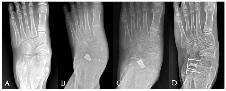
Background/Objectives: Subtalar arthroereisis (STA) is a widely used surgical procedure for symptomatic pediatric flexible flatfoot. However, implant migration remains a concern due to its potential impact on long-term correction and complications. This study evaluated the migration pattern of STA implants and assessed long-term clinical and radiographic outcomes. Methods: This retrospective cohort study included 47 feet from children aged 8-13 years who underwent STA with adjunctive soft tissue procedures between 2014 and 2018, following ≥6 months of failed conservative treatment, with a minimum follow-up of 5 years. Exclusion criteria included neuromuscular or rigid flatfoot. Weight-bearing radiographs assessed anteroposterior (AP) and lateral Meary's angles, reflecting forefoot-to-hindfoot alignment, and calcaneal pitch, indicative of longitudinal arch height. Implant migration was recorded and clinical outcomes were measured by the American Orthopedic Foot and Ankle Society (AOFAS) score. Measurements were recorded preoperatively, immediately postoperatively, and at 1 month, 3 months, 6 months, 1 year, and 5 years. Results: Radiographic correction was significant and sustained at 5 years. The AP Meary's angle improved from 13.09° to 5.26° at 1 month and 6.69° at 5 years (p < 0.001); lateral Meary's angle from 9.77° to 4.06° and 4.88° (p < 0.001); and calcaneal pitch from 14.52° to 16.87° and 16.89° (p < 0.001), respectively. AOFAS scores increased from 67.52 to 90.86 at 1 month and 96.33 at 5 years (p < 0.001). Implant migration peaked within the first postoperative month (mean: 3.2 mm on ankle AP view; 3.0 mm on foot AP view) and stabilized thereafter. Four cases of complications included implant dislodgement, subsidence, and persistent sinus tarsi tenderness, which were successfully resolved after appropriate management. No recurrence of deformity was observed. Conclusions: STA implant migration is most pronounced during the first month, likely due to physiological settling as the foot adapts to altered biomechanics. With appropriate implant selection, technique, and follow-up, migration does not compromise long-term correction or outcomes. In general, symptomatic cases can often be managed conservatively prior to implant removal.
Keywords: flexible flatfoot; implant migration; long-term outcomes; pediatric orthopedics; subtalar arthroereisis.
Conflict of interest statement
The funder is the authors’ hospital for this research project. The funder had no role in study design, data collection, analysis, or interpretation, the writing of the manuscript, or the decision to publish the results.
Figures
Similar articles
-
Mid-term Results of Subtalar Arthroereisis with Talar-Fit Implant in Pediatric Flexible Flatfoot and Identifying the Effects of Adjunctive Procedures and Risk Factors for Sinus Tarsi Pain.Orthop Surg. 2021 Feb;13(1):175-184. doi: 10.1111/os.12864. Epub 2020 Dec 17. Orthop Surg. 2021. PMID: 33332772 Free PMC article.
-
What Are the Functional, Radiographic, and Survivorship Outcomes of a Modified Cup-cage Technique for Pelvic Discontinuity?Clin Orthop Relat Res. 2024 Dec 1;482(12):2149-2160. doi: 10.1097/CORR.0000000000003186. Epub 2024 Jul 9. Clin Orthop Relat Res. 2024. PMID: 38991223
-
Surgical versus non-surgical interventions for displaced intra-articular calcaneal fractures.Cochrane Database Syst Rev. 2023 Nov 7;11(11):CD008628. doi: 10.1002/14651858.CD008628.pub3. Cochrane Database Syst Rev. 2023. PMID: 37933733 Free PMC article.
-
Does Minimally Invasive Surgery Provide Better Clinical or Radiographic Outcomes Than Open Surgery in the Treatment of Hallux Valgus Deformity? A Systematic Review and Meta-analysis.Clin Orthop Relat Res. 2023 Jun 1;481(6):1143-1155. doi: 10.1097/CORR.0000000000002471. Epub 2022 Nov 4. Clin Orthop Relat Res. 2023. PMID: 36332131 Free PMC article.
-
Effect of different subtalar joint arthroereisis implants on pediatric flexible flatfoot: a finite element analysis.Transl Pediatr. 2025 Jun 27;14(6):1296-1305. doi: 10.21037/tp-2025-352. Epub 2025 Jun 25. Transl Pediatr. 2025. PMID: 40688213 Free PMC article.
References
Grants and funding
LinkOut - more resources
Full Text Sources
