. 2025 Jul 14;13(7):1710.
doi: 10.3390/biomedicines13071710.

Evaluation of Apoptosis and Cytotoxicity Induction Using a Recombinant Newcastle Disease Virus Expressing Human IFN-γ in Human Prostate Cancer Cells In Vitro

Affiliations

Evaluation of Apoptosis and Cytotoxicity Induction Using a Recombinant Newcastle Disease Virus Expressing Human IFN-γ in Human Prostate Cancer Cells In Vitro

Aldo Rojas-Neyra et al. Biomedicines. .

Abstract

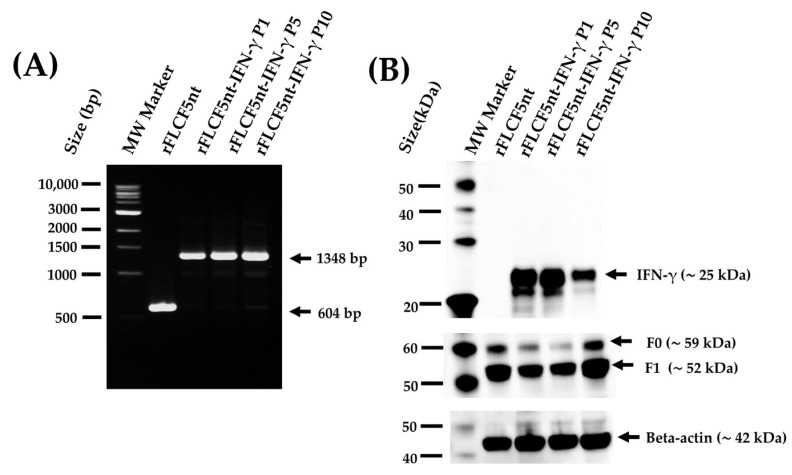
Background/Objectives: Prostate cancer is the second most common type of cancer diagnosed in men. Various treatments for this cancer, such as radiation therapy, surgery, and systemic therapy, can cause side effects in patients; therefore, there is a need to develop new treatment alternatives. One promising approach is virotherapy, which involves using oncolytic viruses (OVs), such as the recombinant Newcastle disease virus (rNDV). Methods: We used the lentogenic rNDV rLS1 strain (the control virus) as our backbone to develop two highly fusogenic rNDVs: rFLCF5nt (the parental virus) and rFLCF5nt-IFN-γ (rFLCF5nt expressing human interferon-gamma (IFN-γ)). We evaluated their oncolytic properties in a prostate cancer cell line (DU145). Results: The results showed the expression and stability of the IFN-γ protein, as confirmed using Western blotting after ten passages in specific pathogen-free chicken embryo eggs using the IFN-γ-expressing virus. Additionally, we detected a significantly high oncolytic activity in DU145 cells infected with the parental virus or the IFN-γ-expressing virus using MTS (a cell viability assay) and Annexin V-PE assays compared with the control virus (p < 0.0001 for both). Conclusions: In conclusion, our data show that IFN-γ-expressing virus can decrease cell viability and induce apoptosis in human prostate cancer in vitro.

Keywords: DU145; Newcastle disease virus; apoptosis; oncolytic.


Conflict of interest statement

The authors are employees from FARVET S.A.C. and declare that this study received funding from FARVET S.A.C. The funder was not involved in the study design, collection, analysis, interpretation of data, the writing of this article or the decision to submit it for publication.

Figures

Figure 1
Figure 1
Characterization of recombinant Newcastle disease viruses. (A) Schematic representation of the generation of the rFLCF5nt and rFLCF5nt-IFN-γ (IFN-γ-expressing virus) viruses, based on modification of the fusion (F) gene cleavage site and insertion of a human IFN-γ expression cassette. (Illustration created with BioRender.com; not drawn to scale). (B) RT-PCR analysis confirming the insertion of the IFN-γ cassette. Amplicon sizes: 604 bp for rLS1 and rFLCF5nt; 1348 bp for rFLCF5nt-IFN-γ. MW: molecular weight marker. (C) Viral replication kinetics of rLS1, rFLCF5nt, and rFLCF5nt-IFN-γ in DU145 cells at an MOI of 0.001, measured using plaque assay. Statistical significance: * p < 0.05, ** p < 0.01, *** p < 0.001, **** p < 0.0001.
Figure 2
Figure 2
Detection of IFN-γ expression in cells infected with recombinant viruses. (A) Immunofluorescence images of DF-1 cells infected with the parental virus (rFLCF5nt) or the IFN-γ-expressing virus (rFLCF5nt-IFN-γ). IFN-γ (red) and NDV proteins (green) were visualized by specific antibody staining; nuclei were counterstained with DAPI (blue). Scale bar: 100 μm. (B) Western blot analysis of DF-1 cell lysates showing IFN-γ expression (~25 kDa), NDV fusion proteins (F0 and F1), and beta-actin as loading control. (C) Quantification of IFN-γ secretion in DU145 cell culture supernatants by ELISA at 24, 48, and 72 h post-infection. Data represent the median ± SD of two independent experiments. Statistical significance: * p < 0.05, ** p < 0.01.
Figure 3
Figure 3
Genetic stability of the IFN-γ-expressing recombinant virus (rFLCF5nt-IFN-γ). (A) RT-PCR analysis showing maintenance of the IFN-γ expression cassette across virus passages 1, 5, and 10 in embryonated chicken eggs. The expected amplicon size (1348 bp) is indicated. MW: molecular weight marker. (B) Western blot analysis confirming stable IFN-γ protein expression (~25 kDa) in DF-1 cells infected with rFLCF5nt-IFN-γ virus from passages 1, 5, and 10. NDV fusion proteins (F0 and F1) and beta-actin were detected as controls.
Figure 4
Figure 4
Evaluation of fusogenic activity induced by recombinant viruses. (A) Representative immunofluorescence images showing syncytium formation in DU145 cells infected with the parental virus (rFLCF5nt), the IFN-γ-expressing virus (rFLCF5nt-IFN-γ), or the control virus (rLS1). NDV proteins are stained in green; nuclei are counterstained with DAPI (blue). Scale bar: 100 μm. (B,C) Quantification of fusion index and syncytium size based on 24 randomly selected fusion areas. (D,E) Plaque size comparison in DF-1 cells infected with the indicated viruses. Data represent mean ± SD of three independent experiments. Statistical significance: * p < 0.05, **** p < 0.0001.
Figure 5
Figure 5
Oncolytic effects of recombinant viruses on DU145 prostate cancer cells. (A) Cell viability measurement by MTS assay at 48 and 72 h post-infection (MOIs of 0.01, 0.1, and 1). (B) Quantification of apoptotic cells after infection with the parental virus (rFLCF5nt), the IFN-γ-expressing virus (rFLCF5nt-IFN-γ), or control virus (rLS1). (C) Representative flow cytometry plots showing apoptosis and necrosis at 48 and 72 h post-infection (hpi). Data represent mean ± SD of triplicate experiments. Statistical significance compared with non-infected cells: *** p < 0.001, **** p < 0.0001.
Figure 6
Figure 6
Chromatin condensation induced by recombinant viruses in DU145 cells. Representative fluorescence microscopy images of DU145 cells infected with the parental virus (rFLCF5nt), the IFN-γ-expressing virus (rFLCF5nt-IFN-γ), or the control virus (rLS1). Nuclei were stained with DAPI (blue). Chromatin condensation, indicative of apoptosis, is highlighted with dotted circles. Scale bar: 100 μm.

References

    1. World Health Organization. [(accessed on 5 October 2022)]. Available online: https://www.who.int/es/news-room/fact-sheets/detail/cancer.
    1. Rawla P. Epidemiology of Prostate Cancer. World J. Oncol. 2019;10:63–89. doi: 10.14740/wjon1191. - DOI - PMC - PubMed
    1. Society American Cancer Cancer Treatment & Survivorship Facts & Figures 2016–2017. [(accessed on 29 March 2023)]. Available online: https://www.cancer.org/content/dam/cancer-org/research/cancer-facts-and-....
    1. Schirrmacher V., van Gool S., Stuecker W. Breaking Therapy Resistance: An Update on Oncolytic Newcastle Disease Virus for Improvements of Cancer Therapy. Biomedicines. 2019;7:66. doi: 10.3390/biomedicines7030066. - DOI - PMC - PubMed
    1. Huang F., Dai C., Zhang Y., Zhao Y., Wang Y., Ru G. Development of Molecular Mechanisms and Their Application on Oncolytic Newcastle Disease Virus in Cancer Therapy. Front. Mol. Biosci. 2022;9:889403. doi: 10.3389/fmolb.2022.889403. - DOI - PMC - PubMed

LinkOut - more resources