Review
. 2025 Jul 15;13(7):1728.
doi: 10.3390/biomedicines13071728.

Cerebral Edema in Traumatic Brain Injury

Affiliations
Review

Cerebral Edema in Traumatic Brain Injury

Santiago Cardona-Collazos et al. Biomedicines. .

Abstract

Cerebral edema is the abnormal accumulation of fluid in any of the tissue compartments of the cerebral parenchyma. It remains a significant challenge in neurotrauma care because it contributes to secondary brain injury, affecting prognosis. This review analyzes the recent literature, including foundational studies, to describe the mechanisms of distinct types of cerebral edema following traumatic brain injury (TBI). Emerging concepts, such as the role of the glymphatic system and heme-derived inflammasomes, offer new insights into new types of edemas, differentiated by pathogenesis and potential treatments. Recent advancements in understanding these molecular mechanisms can improve therapeutic strategies, facilitating a better approach in the era of precision and personalized medicine. Although there has been notable progress, a proposal to customize treatments for diverse types of edemas is necessary to improve outcomes following traumatic brain injury. In this review, we describe the current subtypes of post-traumatic brain edemas and link them to a specific management approach.

Keywords: aquaporins; blood–brain barrier; cerebral edema; glymphatic system; intracranial pressure; traumatic brain injury.


Conflict of interest statement

The authors declare no conflicts of interest.

Figures

Figure 1
Figure 1
Representation of cerebral cytotoxic edema. Cytotoxic edema occurs due to the intracellular accumulation of sodium and water in astrocytes and neurons due to dysfunction of the Na+/K+-ATPase pump during ischemic events. Aquaporins, especially AQP4, facilitate water transport, exacerbating edema and contributing to cellular damage. The red crosses indicate dysfunction of the Na+/K+-ATPase pump, which prevents Na+ from leaving the cell. The black arrow within the AQP4 channel shows the passive influx of water into the cell due to cytotoxic edema.
Figure 2
Figure 2
Representation of ionic edema. This type of edema arises from ionic imbalance due to cytotoxic edema, where ions accumulate inside cells. This creates an osmotic gradient that shifts fluid from the intravascular to the extracellular space across a functional BBB. ① illustrates the osmotic balance among the intravascular, extracellular, and intracellular compartments following the development of cytotoxic edema. Note the high concentration of Na+ and H2O within the intracellular space compared to the extracellular space. ② shows the subsequent movement of Na+ and water from the intravascular compartment into the extracellular space, triggered by the previously described imbalance, leading to the formation of ionic edema. The red crosses indicate dysfunction of the Na+/K+-ATPase pump. The black arrow within the AQP4 channel represents the passive influx of water into the cell due to cytotoxic edema. The black arrows extending from the intravascular to the extracellular space illustrate passive water movement into the extracellular compartment, as occurs in ionic edema.
Figure 3
Figure 3
The images above show CT scans of a patient who presented with multiple frontal contusions and a left occipital epidural hematoma. The patient received conservative management, as determined by the clinical judgment of the attending neurosurgeon (who is not among the authors). The decision was based on two main factors: the patient’s Glasgow Coma Scale score consistently remained at 15, and serial imaging over the first seven days showed that the hematoma size was stable. These images illustrate the chronological evolution of the brain contusions and the progression of distinct types of associated cerebral edema. (A) shows early bifrontal cerebral contusions accompanied by cytotoxic edema. (B) displays the same contusions at a later stage, with increased surrounding edema, reflecting a combination of ongoing cytotoxic edema and the emergence of ionic, vasogenic, and hemolytic edema (described in detail below). (C), taken seven days post-injury, shows more pronounced edema, now predominantly vasogenic in nature. Yellow arrows highlight areas of edema in each panel.
Figure 4
Figure 4
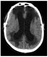
CT imaging shows a combination of ventriculomegaly and periventricular hypodensity characteristic of interstitial edema.
Figure 5
Figure 5
Virchow–Robin spaces: perivascular, fluid-filled canals surrounding perforating brain arteries and veins.
Figure 6
Figure 6
CSF-shift edema after subarachnoid hemorrhage (SAH). Blood in the subarachnoid space and basal cisterns causes a sudden rise in pressure in the subarachnoid compartment. Glymphatic impairment causes CSF to shift from the cerebral cisterns to the brain, leading to swelling. The red crosses represent the inability of interstitial fluid to drain into the venous system, a key aspect of the pathophysiology of CSF-shift edema.
Figure 7
Figure 7
CT scan showing a thick SAH in the perimesencephalic, interhemispheric, and sylvian cisterns (red arrows), accompanied by an acute right-sided subdural hematoma (yellow arrow). Note the underlying CSF-shift edema within the adjacent white matter (blue arrows).
Figure 8
Figure 8
Representation of hemolytic edema pathophysiology. Around the intracerebral hematoma, in the interstitial compartment, hemolytic edema occurs (represented by blue dots) due to the degradation of hemoglobin (red dots). This process triggers different events: First, hemoglobin turns into methemoglobin, releasing iron, carbon monoxide, and biliverdin, which turns into bilirubin by biliverdin reductase. Second, the accumulation of released iron leads to oxidative stress and the formation of free radicals and reactive oxygen species (ROS). Third, the activation of microglia and macrophages stimulates the secretion of tumor necrosis factor (TNF) and the activation of NF-kB, which results in neuroinflammation and necroptosis processes.
Figure 9
Figure 9
CT imaging of an acute subdural hematoma and posterior development of hemolytic cerebral edema. (A) demonstrates an acute subdural hematoma, few hours after injury, with a thickness of less than 10 mm and a midline deviation of less than 5 mm. (B) demonstrates the formation of cerebral edema in the area underlying the hematoma few days after the injury. (C) demonstrates the formation of more cerebral edema that required decompressive craniectomy. The yellow arrow indicates hemolytic edema.
Figure 10
Figure 10
Types of cerebral edema and their distribution in various cerebral compartments.

References

    1. Iencean S.M. Brain edema—A new classification. Med. Hypotheses. 2003;61:106–109. doi: 10.1016/S0306-9877(03)00127-0. - DOI - PubMed
    1. Ho M.L., Rojas R., Eisenberg R.L. Cerebral Edema. Am. J. Roentgenol. 2012;199:W258–W273. doi: 10.2214/AJR.11.8081. - DOI - PubMed
    1. Marmarou A. A review of progress in understanding the pathophysiology and treatment of brain edema. Neurosurg. Focus. 2007;22:1–10. doi: 10.3171/foc.2007.22.5.2. - DOI - PubMed
    1. Stokum J.A., Gerzanich V., Simard J.M. Molecular pathophysiology of cerebral edema. J. Cereb. Blood Flow Metab. 2016;36:513–538. doi: 10.1177/0271678X15617172. - DOI - PMC - PubMed
    1. Jha R.M., Kochanek P.M., Simard J.M. Pathophysiology and treatment of cerebral edema in traumatic brain injury. Neuropharmacology. 2019;145:230–246. doi: 10.1016/j.neuropharm.2018.08.004. - DOI - PMC - PubMed

LinkOut - more resources