. 2025 Jun 28;14(7):803.
doi: 10.3390/antiox14070803.

Unveiling the Cardioprotective Potential of Hydroxytyrosol: Insights from an Acute Myocardial Infarction Model

Affiliations

Unveiling the Cardioprotective Potential of Hydroxytyrosol: Insights from an Acute Myocardial Infarction Model

Alejandra Bermúdez-Oria et al. Antioxidants (Basel). .

Abstract

Cardiovascular diseases remain the leading cause of death worldwide, highlighting the urgent need for novel therapeutic strategies. The Mediterranean diet is renowned for its cardiovascular benefits, largely attributed to extra virgin olive oil (EVOO) and its phenolic compounds, particularly hydroxytyrosol (HT). HT, a potent antioxidant and anti-inflammatory agent, has demonstrated significant therapeutic potential in mitigating myocardial damage following acute myocardial infarction (AMI). However, there is a notable lack of published evidence regarding the effects of HT administration in the context of acute ischemia/reperfusion (I/R) injury, making this study a novel contribution to the field. This study aimed to evaluate the cardioprotective effects of HT using the Langendorff technique in an isolated mouse heart ischemia/reperfusion (I/R) model. Mice were administered a single intraperitoneal dose of HT (10 mg/kg) 24 h prior to the I/R protocols, and parameters such as the infarct size, mitochondrial function, and redox balance were assessed. The results revealed a remarkable 57% reduction in infarct size in HT-treated mice compared to untreated controls. HT treatment also improved mitochondrial bioenergetics, as evidenced by the increased membrane potential (ΔΨm), enhanced oxygen consumption, and reduced hydrogen peroxide (H2O2) production. Furthermore, HT restored the activity of the mitochondrial respiratory complexes, notably Complex I, even under I/R conditions. These findings highlight the efficacy of HT in reducing oxidative stress and preserving mitochondrial function, critical factors in cardiac disease. In conclusion, HT emerges as a promising therapeutic agent for ischemic heart disease, demonstrating both preventive and restorative potential. Future research should explore its clinical applicability to advance cardiovascular disease management.

Keywords: antioxidant; by-products; cardioprotection; hydroxytyrosol (HT); ischemic heart disease (IHD); mitochondrial function.


Conflict of interest statement

The authors declare no conflicts of interest.

Figures

Figure 1
Figure 1
Schematic representation of the isolated heart Langendorff perfusion system and ischemia/reperfusion protocol. This diagram illustrates the experimental setup for maintaining isolated mouse hearts under controlled perfusion conditions. Key components include a thermostatic bath for temperature regulation, a perfusion pump for precise buffer delivery, and a pacemaker/pressure transducer to monitor cardiac function. The system ensures constant retrograde perfusion of the coronary arteries with oxygenated Krebs–Henseleit buffer. Hearts underwent a 15 min stabilization period, followed by 30 min of global ischemia, and then 120 min of reperfusion.
Figure 2
Figure 2
Infarct size, measured as a percentage of the total left ventricular area, was assessed following ischemia/reperfusion (I/R) injury using the triphenyltetrazolium technique. Pre-treatment with HT (10 mg/kg) 24 h prior to I/R resulted in a significant reduction in infarct size compared to the I/R group. For the I/R protocols, ischemia was induced for 30 min followed by 120 min of reperfusion. Data are presented as mean ± standard error of the mean (SEM). Statistical significance is indicated as follows: p * < 0.001 vs. I/R (n I/R group = 7; n HT + I/R group = 4). An independent Student’s t-test was used for comparisons between groups. Representative images of TTC-stained heart sections. Infarct area (pale) is clearly distinguishable from viable myocardium (red).
Figure 3
Figure 3
(a) Representative traces obtained from Clark-type oxygen electrode measurements illustrating μM oxygen consumption over time for the four experimental groups: control, I/R, HT, and HT + I/R. Each trace displays state 4 respiration (basal oxygen consumption) and the subsequent increase in oxygen uptake upon ADP addition, leading to state 3 respiration (ADP-stimulated respiration). (b) Mitochondrial function of the left ventricles was evaluated in control (untreated mice), I/R (mice subjected to 30 min of ischemia and 60 min of reperfusion, I/R), HT (mice treated with HT at the specified dose 24 h previously with lack of I/R protocol), and HT + I/R (mice treated with HT at the specified dose 24 h previously and subjected to I/R). The state 4 and state 3 mitochondrial oxygen consumption rates were evaluated. In addition, the respiratory control (RC) rate was calculated as the state 3/state 4 ratio of the respiratory rate. For the I/R protocols, ischemia was induced for 30 min followed by 60 min of reperfusion. No significant alterations were observed in the respiration in state 4, or resting respiration, between the tested groups. However, in the HT + I/R group, there was a significant increase in active oxygen consumption–state 3–as compared to the I/R group. It should be noted that the I/R group suffered a 54% decrease in O2 consumption values in state 3 as compared to the control (p < 0.01). On the contrary, the HT + I/R group exhibited state 3 O2 consumption levels comparable to those obtained for the control and HT groups. Data are presented as mean ± standard error of the mean (SEM). Statistical significance is indicated as follows: * p < 0.01 indicates a significant effect across groups in the overall analysis; # p < 0.001 vs. control (n control = 3; n I/R group = 3; n HT = 2; n HT + I/R group = 9).
Figure 4
Figure 4
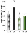
Hydrogen peroxide (H2O2) production measured using the Amplex Red assay. The figure shows H2O2 production rates in the experimental groups: control (untreated mice), I/R (mice subjected to I/R), HT (mice treated with HT at the specified dose), and HT + I/R (mice treated with HT at the specified dose and subjected to I/R). For the I/R protocols, ischemia was induced for 30 min followed by 60 min of reperfusion. The I/R group exhibited the highest H2O2 production as compared to controls. Notably, the HT + I/R group showed lower production levels as compared to the control group. Furthermore, the HT group displayed the lowest H2O2 production. Data are presented as mean ± standard error of the mean (SEM). Statistical significance is indicated as follows: * p < 0.05 indicates a significant effect across groups in the overall analysis; # p < 0.001 vs. control; p < 0.001 vs. I/R (n control = 3; n I/R group = 5; n HT = 9; n HT + I/R group = 11).
Figure 5
Figure 5
Mitochondrial membrane potential (ΔΨm) in state 4 respiration measured using the rhodamine assay. The figure shows ΔΨm in four experimental groups: control (untreated mice), I/R (mice subjected to I/R), HT (mice treated with HT at the specified dose), and HT + I/R (mice treated with HT at the specified dose and subjected to I/R). For the I/R protocols, ischemia was induced for 30 min followed by 60 min of reperfusion. It should be noted that the HT + I/R group maintained ΔΨ levels comparable to those of the control group, in contrast to the decrease of about 10% observed in the I/R group. In accordance with the previous findings, the HT group once again showed significantly higher ΔΨ values (193 ± 5), thus indicating a protective effect of HT upon mitochondrial coupling in basal conditions. Data are presented as mean ± standard error of the mean (SEM). Statistical significance is indicated as follows: * p < 0.05 indicates a significant effect across groups in the overall analysis; # p < 0.001 vs. control;  p < 0.001 vs. I/R (n control = 3; n I/R group = 5; n HT = 4; n HT + I/R group = 4).
Figure 6
Figure 6
Activity of mitochondrial Complex I in the left ventricle of mice subjected to isolated heart I/R. The figure displays Complex I activity across four experimental groups: control (untreated mice), I/R (mice subjected to I/R), HT (mice treated with HT at the specified dose), and HT + I/R (mice treated with HT at the specified dose and subjected to I/R). For the I/R protocols, ischemia was induced for 30 min followed by 60 min of reperfusion. A notable increase in this activity was detected in the HT + I/R group compared to the I/R group, reaching levels similar to those of the control group. Treatment with HT administered 24 h prior to euthanasia resulted in a significant increase in Complex I activity in the group not subjected to the I/R protocol. Data are presented as mean ± standard error of the mean (SEM). Statistical significance is indicated as follows: * p < 0.05 indicates a significant effect across groups in the overall analysis; # p < 0.001 vs. control;  p < 0.001 vs. I/R (n control =3; n I/R group =3; n HT= 5; n HT + I/R group = 10).
Figure 7
Figure 7
Proposed mechanism of hydroxytyrosol (HT) cardioprotection against ischemia/reperfusion (I/R) injury. Hydroxytyrosol (HT) acts within the cardiomyocyte to mitigate I/R damage. Upon entering the cell, HT activates the Nrf2 pathway, leading to its nuclear translocation and subsequent increased expression of antioxidant genes (ARE). The enhanced antioxidant defense system, along with HT’s direct effects, results in reduced oxidative stress (decreased H2O2). This action, coupled with improved mitochondrial function (evidenced by increased respiration, enhanced mitochondrial potential, and improved mitochondrial complex activity), collectively leads to cardioprotection and a reduction in myocardial infarct size.

Similar articles

References

    1. AL-Asmari1 K.M., Al-Attar A.M., Abu Zeid I.M. Potential health benefits and components of oliveoil: An overview. Biosci. Res. 2020;17:2673–2687.
    1. Guasch-Ferré M., Hu F.B., Martínez-González M.A., Fitó M., Bulló M., Estruch R., Ros E., Corella D., Recondo J., Gómez-Gracia E., et al. Olive oil intake and risk of cardiovascular disease and mortality in the PREDIMED Study. BMC Med. 2024;12:78. doi: 10.1186/1741-7015-12-78. - DOI - PMC - PubMed
    1. Di Cesare M., Perel P., Taylor S., Kabudula C., Bixby H., Gaziano T.A., McGhie D.V., Mwangi J., Pervan B., Narula J., et al. The Heart of the World. Glob. Heart. 2024;19:11. doi: 10.5334/gh.1288. - DOI - PMC - PubMed
    1. Neumann F.-J., Sousa-Uva M., Ahlsson A., Alfonso F., Banning A.P., Benedetto U., Byrne R.A., Collet J.-P., Falk V., Head S.J., et al. ESC/EACTS Guidelines on Myocardial Revascularization. Eur. Heart J. 2018;40:87–165. doi: 10.1093/eurheartj/ehy394. - DOI - PubMed
    1. García-Niño W.R., Zazueta C., Buelna-Chontal M., Silva-Palacios A. Mitochondrial Quality Control in Cardiac-Conditioning Strategies against Ischemia-Reperfusion Injury. Life. 2021;11:1123. doi: 10.3390/life11111123. - DOI - PMC - PubMed

LinkOut - more resources