. 2025 Jun 30;14(7):808.
doi: 10.3390/antiox14070808.

Carnosol, a Rosemary Ingredient Discovered in a Screen for Inhibitors of SARM1-NAD+ Cleavage Activity, Ameliorates Symptoms of Peripheral Neuropathy

Affiliations

Carnosol, a Rosemary Ingredient Discovered in a Screen for Inhibitors of SARM1-NAD+ Cleavage Activity, Ameliorates Symptoms of Peripheral Neuropathy

Hitoshi Murata et al. Antioxidants (Basel). .

Abstract

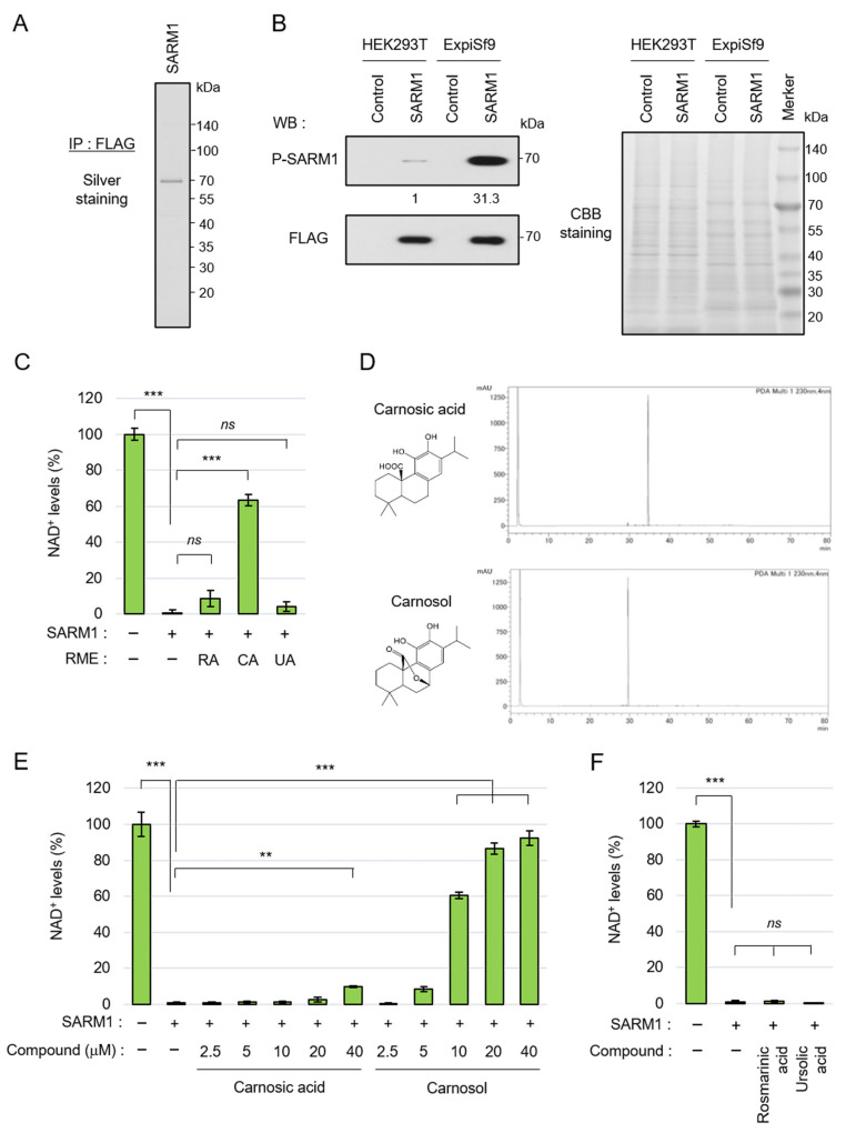
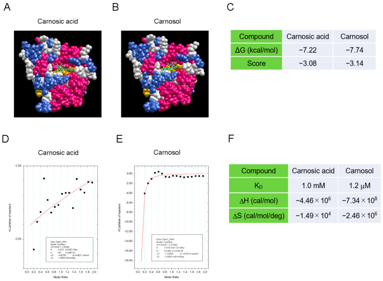
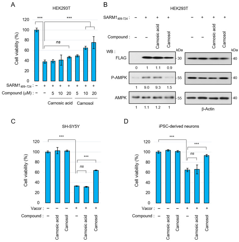
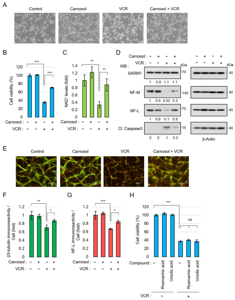
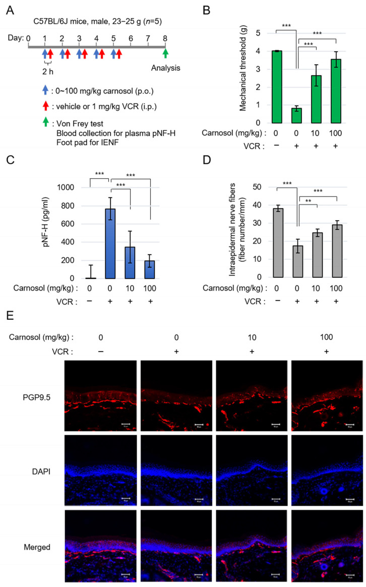
Sterile alpha and Toll/interleukin receptor motif-containing protein 1 (SARM1) is a nicotinamide adenine dinucleotide (NAD+) hydrolase involved in axonal degeneration and neuronal cell death. SARM1 plays a pivotal role in triggering the neurodegenerative processes that underlie peripheral neuropathies, traumatic brain injury, and neurodegenerative diseases. Importantly, SARM1 knockdown or knockout prevents the degeneration; as a result, SARM1 has been attracting attention as a potent therapeutic target. In recent years, the development of several SARM1 inhibitors derived from synthetic chemical compounds has been reported; however, no dietary ingredients with SARM1 inhibitory activity have been identified. Therefore, we here focused on dietary ingredients and found that carnosol, an antioxidant contained in rosemary, inhibits the NAD+-cleavage activity of SARM1. Purified carnosol inhibited the enzymatic activity of SARM1 and suppressed neurite degeneration and cell death induced by the anti-cancer medicine vincristine (VCR). Carnosol also inhibited VCR-induced hyperalgesia symptoms, suppressed the loss of intra-epidermal nerve fibers in vivo, and reduced the blood fluid level of phosphorylated neurofilament-H caused by an axonal degeneration event. These results indicate that carnosol has a neuroprotective effect via SARM1 inhibition in addition to its previously known antioxidant effect via NF-E2-related factor 2 and thus suppresses neurotoxin-induced peripheral neuropathy.

Keywords: NAD+; SARM1; axon degeneration; carnosol; peripheral neuropathy.


Conflict of interest statement

The authors, Hitoshi Murata, Kazuki Ogawa, Yoji Wada, Hiromichi Nakamura, and Masakiyo Sakaguchi have filed a patent application for the research in this article. The remaining authors declare that they have no conflicts of interest with respect to the contents of this article.

Figures

Figure 1
Figure 1
Carnosol inhibited SARM1-NADase activity: (A) silver staining gel of purified SARM1 protein. A full-length SARM1-FLAG protein was purified from lysates of ExpiSf9; (B) comparison of phosphorylation levels of SARM1 expressed in HEK293T and ExpiSf9. The band intensities of P-SARM1 were normalized to that of FLAG and the values are indicated below the panel. A CBB-stained gel of cell lysates was used as a loading control; (C) inhibition analysis of SARM1-NADase using rosemary extracts. A total of 1 μM SARM1 protein was incubated with 10 μg/mL RME–RA, RME–CA, or RME–UA; (D) HPLC data of purified carnosic acid and carnosol from rosemary extracts; (E) inhibition analysis of SARM1-NADase using carnosic acid and carnosol. A total of 1 μM SARM1 protein was incubated with 0~40 μM of the compounds; and (F) rosmarinic acid and ursolic acid did not have SARM1 inhibitory activity. A total of 1 μM SARM1 protein was incubated with 10 μM compounds. ns: not significant; ** p < 0.01; *** p < 0.001.
Figure 2
Figure 2
Interaction between the SARM1-TIR domain and carnosol (AC). An in silico assay of the interaction between the SARM1-TIR domain and compounds by myPresto Portal. The cryo-EM structure of activated human SARM1 in a complex with NMN and 1AD (7NAK; Protein Data Bank) was used as the receptor molecule: (A) carnosic acid (PubChem CID: 65126); (B) carnosol (PubChem CID: 442009) were used as docking compounds of the SARM1-TIR domain; and (C) the docking score of compounds to the SARM1-TIR domain. (D,F) Analysis of the direct interaction between SARM1594–670 and compounds using isothermal titration calorimetry. 20 μM GST-fused SARM1594–670 was used as the receptor molecule: (D) 200 μM carnosic acid; (E) 200 μM carnosol were used as ligands of SARM1594–670; and (F) the ability of the tested compounds to interact with GST-SARM1594–670.
Figure 3
Figure 3
Suppression of SARM1-dependent cytotoxic activity by carnosol: (A) relative cell viability of HEK293T cells expressing SARM1409–724 treated with 0~20 μM carnosic acid or 0~20 μM carnosol for 16 h; (B) carnosol suppressed the AMPK phosphorylation induced by SARM1409–724. HEK293T cells expressing SARM1409–724 were treated with DMSO (control), 10 μM carnosic acid, or 10 μM carnosol for 16 h. The band intensities of the indicated proteins were normalized to their corresponding bands of β-Actin in the right panel and the values are indicated below each panel; (C) suppression of vacor-induced cell death by carnosol in SH-SY5Y cells. SH-SY5Y cells were treated for 8 h with 50 μM vacor, 10 μM carnosic acid, or 10 μM carnosol; and (D) suppression of vacor-induced cell death by carnosol in iPSC-derived neurons. Human iPSC-derived neurons were treated for 8 h with 10 μM vacor, 10 μM carnosic acid, or 10 μM carnosol. ns: not significant; *** p < 0.001.
Figure 4
Figure 4
Carnosol suppressed vincristine (VCR)-induced neurite degeneration and cell death: (AG) human iPSC-derived neurons were treated with 50 nM VCR and 10 μM carnosol for 24 h (AD) or 8 h (EG). Panels (AD) show, as follows: (A) cell morphologies, scale bar: 200 μm; (B) cell viability; (C) NAD+ levels; and (D) Western blotting results for cell lysates at 24 h after the treatment. The band intensities of the indicated proteins were normalized to their corresponding bands of β-Actin in the right panel and the values are indicated below each panel. (E) Representative images of neuronal axons after 8 h of treatment with the indicated compounds. Stain: anti-β3 tubulin and NF-L. Scale bar: 100 μm; (F,G) immunoreactivity of β3-tubulin and NF-L. The fluorescence intensities of β3-tubulin and NF-L were normalized against nuclear numbers; and (H) treatment with rosmarinic acid or ursolic acid did not suppress VCR-induced cell death. The cell viabilities of iPSC-derived neurons that were treated for 24 h with 50 nM VCR, 10 μM rosmarinic acid, or 10 μM ursolic acid. ns: not significant, * p < 0.05, ** p < 0.01, *** p < 0.001.
Figure 5
Figure 5
Carnosol treatment attenuated VCR-induced peripheral neuropathy: (A) schematic diagram of the treatment. (B) quantification of mechanical sensitivity; (C) quantification of plasma pNF-H levels; and (D,E) the changes in INEF density in mice given the treatment. Quantification data (D) and the representative figures (E). Stain: anti-PGP9.5 and DAPI. Scale bar: 50 μm. ** p < 0.01, *** p < 0.001.

Similar articles

References

    1. Coleman M.P., Hoke A. Programmed axon degeneration: From mouse to mechanism to medicine. Nat. Rev. Neurosci. 2020;21:183–196. doi: 10.1038/s41583-020-0269-3. - DOI - PMC - PubMed
    1. Salvadores N., Geronimo-Olvera C., Court F.A. Axonal Degeneration in AD: The Contribution of Abeta and Tau. Front. Aging Neurosci. 2020;12:581767. doi: 10.3389/fnagi.2020.581767. - DOI - PMC - PubMed
    1. Burke R.E., O’Malley K. Axon degeneration in Parkinson’s disease. Exp. Neurol. 2013;246:72–83. doi: 10.1016/j.expneurol.2012.01.011. - DOI - PMC - PubMed
    1. Chaudhry V., Cornblath D.R. Wallerian degeneration in human nerves: Serial electrophysiological studies. Muscle Nerve. 1992;15:687–693. doi: 10.1002/mus.880150610. - DOI - PubMed
    1. Wang M.S., Wu Y., Culver D.G., Glass J.D. Pathogenesis of axonal degeneration: Parallels between Wallerian degeneration and vincristine neuropathy. J. Neuropathol. Exp. Neurol. 2000;59:599–606. doi: 10.1093/jnen/59.7.599. - DOI - PubMed

LinkOut - more resources