Review
. 2025 Jul 8;14(7):836.
doi: 10.3390/antiox14070836.

Modulation of Redox-Sensitive Cardiac Ion Channels

Affiliations
Review

Modulation of Redox-Sensitive Cardiac Ion Channels

Razan Orfali et al. Antioxidants (Basel). .

Abstract

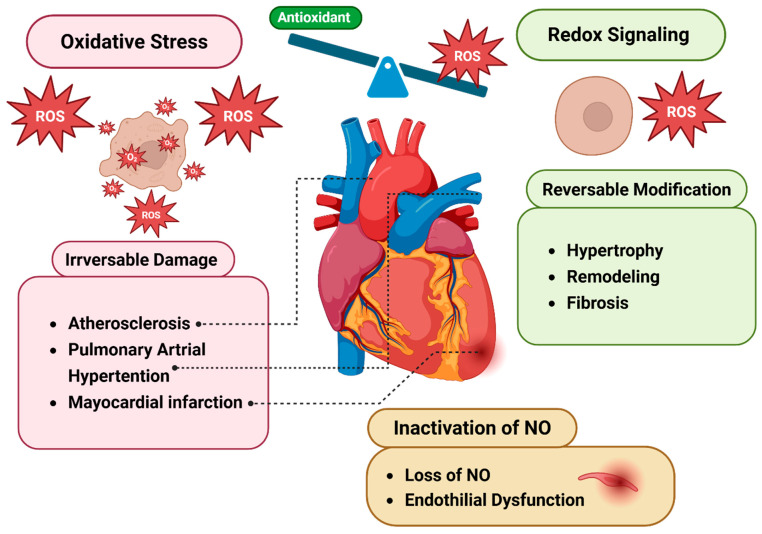
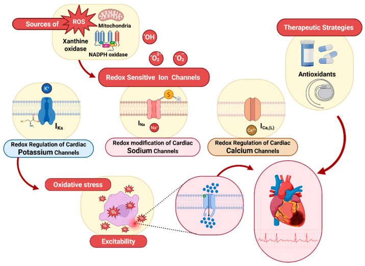
Redox regulation is crucial for the cardiac action potential, coordinating the sodium-driven depolarization, calcium-mediated plateau formation, and potassium-dependent repolarization processes required for proper heart function. Under physiological conditions, low-level reactive oxygen species (ROS), generated by mitochondria and membrane oxidases, adjust ion channel function and support excitation-contraction coupling. However, when ROS accumulate, they modify a variety of important channel proteins in cardiomyocytes, which commonly results in reducing potassium currents, enhancing sodium and calcium influx, and enhancing intracellular calcium release. These redox-driven alterations disrupt the cardiac rhythm, promote after-depolarizations, impair contractile force, and accelerate the development of heart diseases. Experimental models demonstrate that oxidizing agents reduce repolarizing currents, whereas reducing systems restore normal channel activity. Similarly, oxidative modifications of calcium-handling proteins amplify sarcoplasmic reticulum release and diastolic calcium leak. Understanding the precise redox-dependent modifications of cardiac ion channels would guide new possibilities for targeted therapies aimed at restoring electrophysiological homeostasis under oxidative stress, potentially alleviating myocardial infarction and cardiovascular dysfunction.

Keywords: antioxidants; cardiomyocytes; cardiovascular diseases; heart failure; ion channels; kinase; mitochondria; oxidative stress; reactive oxygen species; redox signaling.


Conflict of interest statement

The authors declare no conflicts of interest.

Figures

Figure 1
Figure 1
Simplified ventricular myocyte showing action-potential phases and ion channel localization. Connexin43-based gap junctions, Igi (green) at intercellular borders, conduct rapid ionic currents between adjacent myocytes, triggering Phase 0 depolarization. This activation opens fast inward currents (red), primarily INa and ICa,T, which inactivate quickly, while L-type Ca2+ channels (ICa,L) remain active longer to sustain the plateau (Phase 2). The depolarizing influence of ICa,L is then opposed by early outward K+ currents (Ito, blue), and by ultrarapid/delayed rectifier currents (IKur, IKr, IKs), which drive repolarization. Finally, inward rectifier K+ current (IK1, dark blue) restores the membrane to its resting potential (approximately −85 mV) [25]. Phase numbers (0–4) are marked along the voltage trace.
Figure 2
Figure 2
Primary pathways of reactive oxygen species (ROS) generation in cardiomyocytes. Within mitochondria, a small portion of electrons “leaks” from complexes I, II, and III onto O2, forming the superoxide anion (O2·). Superoxide also arises in the cytosol when NADPH oxidase transfers electrons from NADPH to O2 and when xanthine oxidase oxidizes hypoxanthine to xanthine. Mitochondrial O2· diffuses into the cytosol, where it can convert hydrogen peroxide (H2O2) into hydroxyl radicals (·OH) via the Haber–Weiss reaction. Peroxisomes generate much of the cell’s H2O2, but if peroxisomal defenses fail, H2O2 spills into the cytosol, fueling further ·OH production. Under metabolic stress, iron released from 4Fe_4S enzyme clusters drives the Fenton reaction, producing additional ·OH from H2O2. Lipid peroxidation creates membrane peroxyl radicals (ROO·), while excessive nitric oxide (·NO), formed by nitric oxide synthase, combines with O2· to yield the potent oxidant peroxynitrite (ONOO), contributing to nitrosative stress [13].
Figure 3
Figure 3
Antioxidant systems in cardiomyocytes. Key antioxidants include reduced glutathione (GSH) and its oxidized form (GSSG), supported by antioxidant enzymes such as glutathione reductase (GRed), glutathione peroxidase (GPx), catalase, and superoxide dismutase (SOD). Additional components include NADPH oxidase, α-tocopherol (vitamin E), and vitamin C. Abbreviations: (RCOO) lipid radical; (RCOOH) lipid; (α-toco) α-tocopheroxyl radical; (α-toco) α-tocopherol.
Figure 4
Figure 4
The impact of oxidative stress on heart diseases. An imbalance between ROS and antioxidants shifts redox signaling toward oxidative stress, contributing to heart failure, hypertension, and atherosclerosis, often resulting in irreversible cardiac damage. ROS, reactive oxygen species; NO, nitric oxide [73].
Figure 5
Figure 5
Redox-dependent modulation of a cardiac ion channel gating. Reactive oxygen species (ROS) oxidize critical cysteine thiols (orange) on the voltage-sensing domain of the voltage-gated channel, converting them into sulfenic and disulfide forms (green). In the left panel, ROS (red burst) targets the resting channel, initiating thiol oxidation and a partial opening state. These oxidative modifications shift the activation threshold and increase open probability, as depicted on the right by more frequent and longer-lasting ion flux (dense blue dots). Under reducing conditions, thiol groups are restored, and normal gating is recovered.
Figure 6
Figure 6
Diagram of redox-dependent modulation of cardiac ion channels and therapeutic interventions. Mitochondrial electron-transport chain leakage, NADPH oxidases, and xanthine oxidase generate reactive oxygen species (ROS: O2, H2O2, •OH), which impart redox modifications (e.g., S-nitrosylation, glutathionylation, disulfide formation) on voltage-gated Na+, Ca2+, and K+ channels. These thiol-based modifications alter channel gating, leading to disrupted excitability and contractility in ventricular myocytes. Targeted therapeutic strategies, including antioxidants that boost endogenous reductive systems, soluble guanylate cyclase (sGC) stimulators that enhance cGMP-PKG–mediated thiol protection, and device-based pacing (e.g., LBBAP), aim to reverse maladaptive channel oxidation, restore electrical stability, and improve contractile function in oxidative cardiac disease.

References

    1. Uttara B., Singh A.V., Zamboni P., Mahajan R.T. Oxidative Stress and Neurodegenerative Diseases: A Review of Upstream and Downstream Antioxidant Therapeutic Options. Curr. Neuropharmacol. 2009;7:65–74. doi: 10.2174/157015909787602823. - DOI - PMC - PubMed
    1. Mandal M., Sarkar M., Khan A., Biswas M., Masi A., Rakwal R., Agrawal G.K., Srivastava A., Sarkar A. Reactive Oxygen Species (ROS) and Reactive Nitrogen Species (RNS) in plants—Maintenance of structural individuality and functional blend. Adv. Redox Res. 2022;5:100039. doi: 10.1016/j.arres.2022.100039. - DOI
    1. Juan C.A., Pérez de la Lastra J.M., Plou F.J., Pérez-Lebeña E. The Chemistry of Reactive Oxygen Species (ROS) Revisited: Outlining Their Role in Biological Macromolecules (DNA, Lipids and Proteins) and Induced Pathologies. Int. J. Mol. Sci. 2021;22:4642. doi: 10.3390/ijms22094642. - DOI - PMC - PubMed
    1. Bhatti J.S., Bhatti G.K., Reddy P.H. Mitochondrial dysfunction and oxidative stress in metabolic disorders—A step towards mitochondria based therapeutic strategies. Biochim. Biophys. Acta (BBA)—Mol. Basis Dis. 2017;1863:1066–1077. doi: 10.1016/j.bbadis.2016.11.010. - DOI - PMC - PubMed
    1. Kozlov A.V., Javadov S., Sommer N. Cellular ROS and Antioxidants: Physiological and Pathological Role. Antioxidants. 2024;13:602. doi: 10.3390/antiox13050602. - DOI - PMC - PubMed

LinkOut - more resources