. 2025 Jul 14;14(7):859.
doi: 10.3390/antiox14070859.

Negative Air Ions Attenuate Nicotine-Induced Vascular Endothelial Dysfunction by Suppressing AP1-Mediated FN1 and SPP1

Affiliations

Negative Air Ions Attenuate Nicotine-Induced Vascular Endothelial Dysfunction by Suppressing AP1-Mediated FN1 and SPP1

Sha Xiao et al. Antioxidants (Basel). .

Abstract

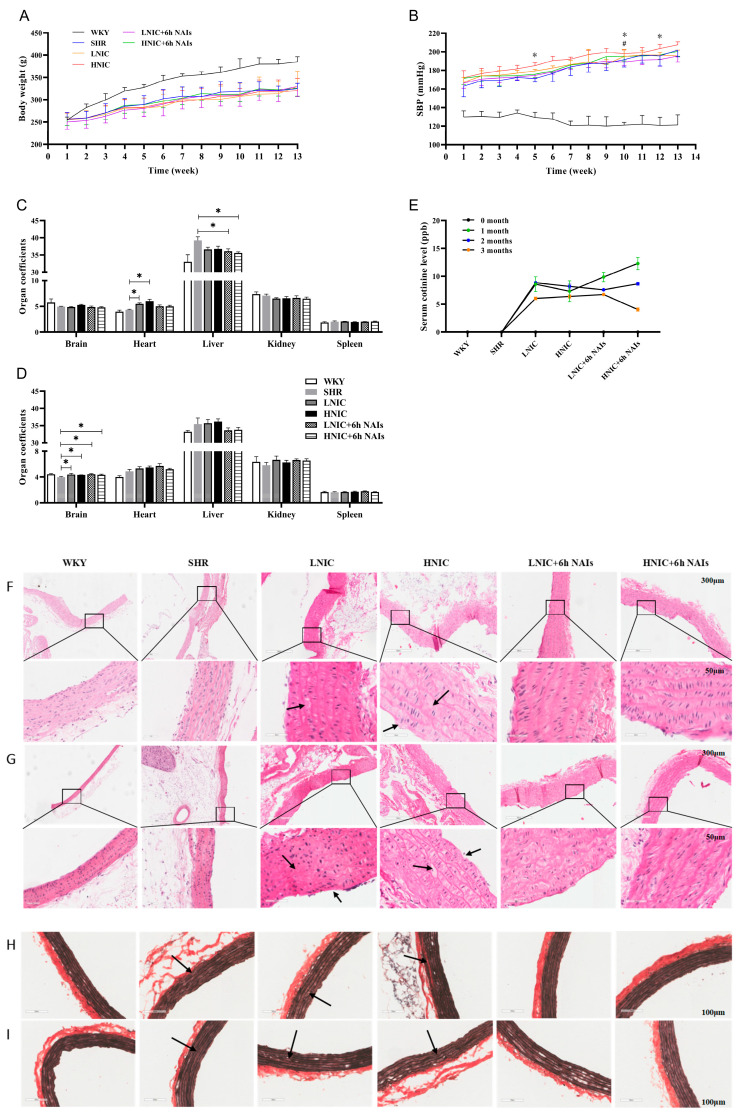
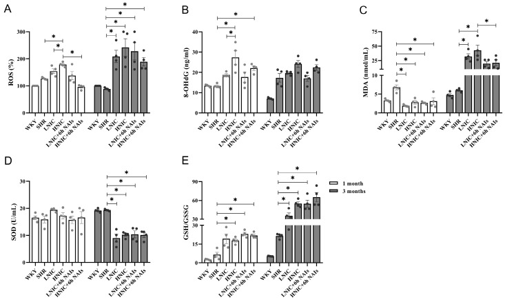
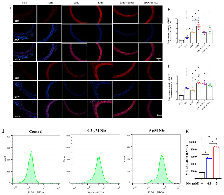
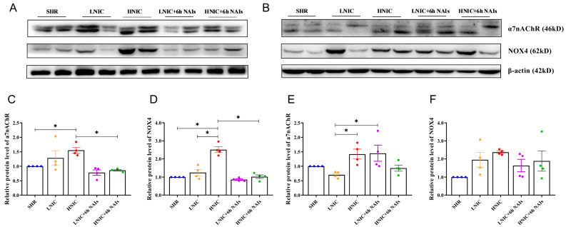
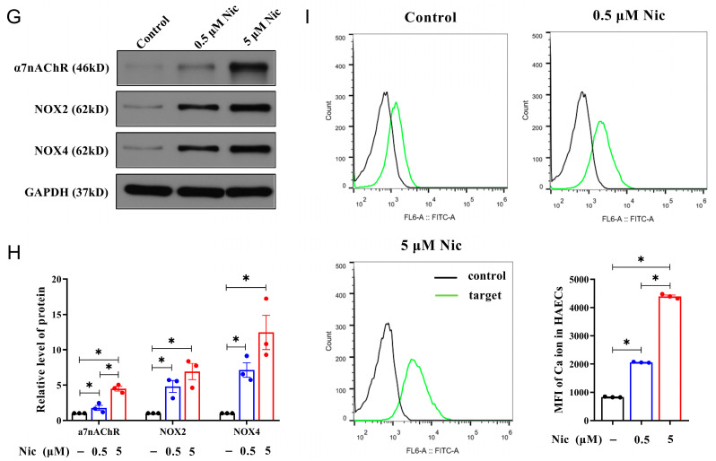
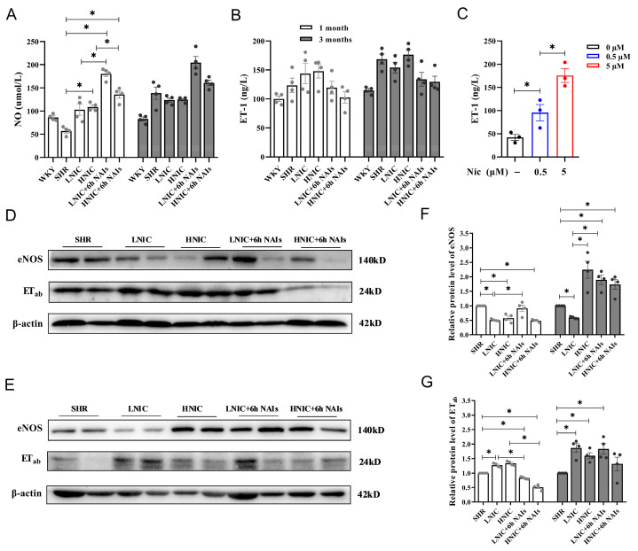
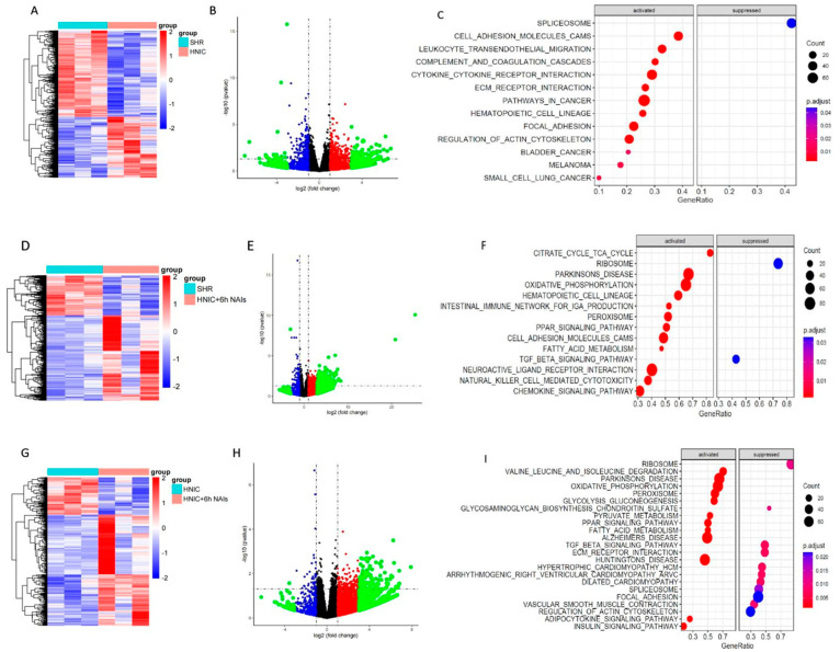
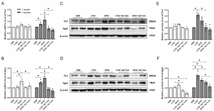
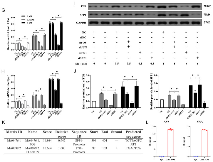
Nicotine-induced oxidative stress contributes significantly to vascular endothelial dysfunction. While negative air ions (NAIs) demonstrate potential blood-pressure-regulating and antioxidant properties, their mechanistic role remains unclear. This study examined the effects of NAIs against nicotine-induced oxidative damage and vascular endothelial injury in spontaneously hypertensive rats (SHRs). Western blotting was used to detect the expression levels of the α7nAChR/MAPK/AP1 pathway. Transcriptomic sequencing was performed to identify the differentially expressed genes after treatment with nicotine or NAIs. Furthermore, reactive oxygen species (ROS), endothelin-1 (ET-1), and [Ca2+]i levels were detected in human aortic endothelial cells (HAECs) treated with nicotine, and the relationship between transcription factor activator protein 1 (AP1) and the target genes was further elucidated through ChIP-qPCR. Nicotine exposure in SHRs elevated blood pressure and induced oxidative damage through α7nAChR/MAPK/AP1 pathway activation, causing endothelial structural disruption. These effects manifested as decreased NO/eNOS and increased ET-1/ETab expression, while these changes were reversed by NAIs. In HAECs, nicotine impaired proliferation while increasing oxidative stress and [Ca2+]i levels. This endothelial damage was markedly attenuated by either NAIs or fibronectin 1 (Fn1)/secreted phosphoprotein 1 (Spp1) knockdown. Mechanistically, we identified AP1 as the transcriptional regulator of FN1 and SPP1. NAIs attenuate nicotine-induced endothelial dysfunction in hypertension by inhibiting AP1-mediated FN1 and SPP1 activation, providing novel insights for smoking-associated cardiovascular risk.

Keywords: activator protein-1; hypertension; negative air ions; nicotine; vascular endothelial dysfunction.


Conflict of interest statement

The authors declare no conflicts of interest.

Figures

Figure 1
Figure 1
Physiological parameters of rats. (A) Body weight (n = 10). (B) Systolic blood pressure (n = 10), * LNIC + 6h NAIs compared with LNIC, p < 0.05; # HNIC + 6h NAIs compared with HNIC, p < 0.05. Rat organ coefficients (n = 5): (C) 1 month; (D) 3 months. (E) Serum cotinine levels (n = 3). Effects of nicotine exposure and NAI intervention on thoracic aorta pathology in rats. HE staining: (F) 1 month; (G) 3 months. Black arrow: endothelial cell defect, thickening of the middle layer, rupture of the smooth muscle fiber layer, and formation of cavities in smooth muscle cells; rupture of the outer membrane. EVG staining: (H) 1 month; (I) 3 months. Black arrow: the elastic fibers show signs of fracture, loss and degradation. ANOVA, * p < 0.05.
Figure 2
Figure 2
Serum oxidative levels in rats (n = 4). (A) ROS levels. (B) 8-OHdG levels. (C) MDA levels. (D) SOD levels. (E) GSH/GSSG levels. DHE fluorescence staining of thoracic aortas in rats exposed to nicotine (n = 4, 100 μm): (F) 1 month; (G) 3 months. ROS levels in the thoracic aortas of rats: (H) 1 month; (I) 3 months. (J) ROS levels of HAECs treated with nicotine. (K) Fluorescence intensity of ROS concentration in HAECs. p-values were obtained through ANOVA; * p < 0.05.
Figure 2
Figure 2
Serum oxidative levels in rats (n = 4). (A) ROS levels. (B) 8-OHdG levels. (C) MDA levels. (D) SOD levels. (E) GSH/GSSG levels. DHE fluorescence staining of thoracic aortas in rats exposed to nicotine (n = 4, 100 μm): (F) 1 month; (G) 3 months. ROS levels in the thoracic aortas of rats: (H) 1 month; (I) 3 months. (J) ROS levels of HAECs treated with nicotine. (K) Fluorescence intensity of ROS concentration in HAECs. p-values were obtained through ANOVA; * p < 0.05.
Figure 3
Figure 3
Protein expression of α7nAChR and NOX4 in the thoracic aorta of rats (n = 4): (A) 1 month; (B) 3 months. Protein quantification of α7nAChR and NOX4: (C,D) 1 month; (E,F) 3 months. (G) Protein expression profiling of α7nAChR, NOX2, and NOX4 in HAECs. (H) Protein quantification of α7nAChR, NOX2, and NOX4 in HAECs. (I) [Ca2+]i concentration of HAECs treated with nicotine; fluorescence intensity at 488 nm. p value was obtained by ANOVA; * p < 0.05.
Figure 3
Figure 3
Protein expression of α7nAChR and NOX4 in the thoracic aorta of rats (n = 4): (A) 1 month; (B) 3 months. Protein quantification of α7nAChR and NOX4: (C,D) 1 month; (E,F) 3 months. (G) Protein expression profiling of α7nAChR, NOX2, and NOX4 in HAECs. (H) Protein quantification of α7nAChR, NOX2, and NOX4 in HAECs. (I) [Ca2+]i concentration of HAECs treated with nicotine; fluorescence intensity at 488 nm. p value was obtained by ANOVA; * p < 0.05.
Figure 4
Figure 4
Effects of nicotine exposure and NAI intervention on the function of the vascular endothelium in rats (n = 4): (A) Serum NO level. (B) Serum ET-1 levels. (C) Supernatant ET-1 concentration of HAECs treated with nicotine. Protein expression of eNOS and ETab in thoracic aortas that were exposed to nicotine: (D) 1 month; (E) 3 months. (F,G) Protein quantification of eNOS and ETab. p-values were obtained through ANOVA; * p < 0.05.
Figure 5
Figure 5
Nicotine exposure promotes the phosphorylation of JNK and JUN in the thoracic aorta of rats (n = 4). Protein expression profiling of MAPK pathway genes: (A) 1 month; (B) 3 months. Protein quantification of MAPK pathway genes in the thoracic aortas of rats: (C) 1 month; (D) 3 months. Protein expression profiling of JUN in the thoracic aortas of rats: (E) 1 month; (F) 3 months. Protein quantification of JUN in the thoracic aortas of rats: (G) 1 month; (H) 3 months. p-values were obtained through ANOVA; * p < 0.05.
Figure 6
Figure 6
RNA-seq was conducted to explore the regulation of gene expression in the thoracic aorta of rats through nicotine exposure and NAI intervention (n = 3). (AC) HNIC group compared to the SHR group. (DF) HNIC+6h NAI group compared to the SHR group. (GI) HNIC+6h NAI group compared to the HNIC group.
Figure 7
Figure 7
Nicotine upregulates mRNA and protein expression levels of Fn1 and Spp1 in the thoracic aortas of rats (n = 4). (A) mRNA expression profiling of Fn1 in rats. (B) mRNA expression profiling of Spp1 in rats. Protein expression profiling of Fn1 and Spp1 in rats: (C) 1 month; (D) 3 months. Protein quantification of Fn1 and Spp1 in rats: (E) 1 month; (F) 3 months. Nicotine regulates activation of FN1 and SPP1 in HAECs via AP1 (n = 3). (G) mRNA expression profiling of FN1 in HAECs. (H) mRNA expression profiling of SPP1 in HAECs. (I) Protein expression profiling of FN1 and SPP1 in HAECs. (J) Protein quantification of FN1 and SPP1 in HAECs. (K) Candidate putative sequences required for the binding of AP1 to the FN1 or SPP1 gene promoter, predicted by the UCSC database and JASPAR database. (L) ChIP-qPCR data for FOS binding at the FN1 promoter and AP1 binding at the SPP1 promoter in HAECs. p-values were obtained through ANOVA; * p < 0.05.
Figure 7
Figure 7
Nicotine upregulates mRNA and protein expression levels of Fn1 and Spp1 in the thoracic aortas of rats (n = 4). (A) mRNA expression profiling of Fn1 in rats. (B) mRNA expression profiling of Spp1 in rats. Protein expression profiling of Fn1 and Spp1 in rats: (C) 1 month; (D) 3 months. Protein quantification of Fn1 and Spp1 in rats: (E) 1 month; (F) 3 months. Nicotine regulates activation of FN1 and SPP1 in HAECs via AP1 (n = 3). (G) mRNA expression profiling of FN1 in HAECs. (H) mRNA expression profiling of SPP1 in HAECs. (I) Protein expression profiling of FN1 and SPP1 in HAECs. (J) Protein quantification of FN1 and SPP1 in HAECs. (K) Candidate putative sequences required for the binding of AP1 to the FN1 or SPP1 gene promoter, predicted by the UCSC database and JASPAR database. (L) ChIP-qPCR data for FOS binding at the FN1 promoter and AP1 binding at the SPP1 promoter in HAECs. p-values were obtained through ANOVA; * p < 0.05.

Similar articles

References

    1. Liu S., Li Y., Zeng X., Wang H., Yin P., Wang L., Liu Y., Liu J., Qi J., Ran S., et al. Burden of cardiovascular diseases in China, 1990–2016: Findings from the 2016 global burden of disease study. JAMA Cardiol. 2019;4:342–352. doi: 10.1001/jamacardio.2019.0295. - DOI - PMC - PubMed
    1. Münzel T., Hahad O., Kuntic M., Keaney J.F., Deanfield J.E., Daiber A. Effects of tobacco cigarettes, e-cigarettes, and waterpipe smoking on endothelial function and clinical outcomes. Eur. Heart J. 2020;41:4057–4070. doi: 10.1093/eurheartj/ehaa460. - DOI - PMC - PubMed
    1. Whitehead A.K., Erwin A.P., Yue X. Nicotine and vascular dysfunction. Acta Physiol. 2021;231:e13631. doi: 10.1111/apha.13631. - DOI - PMC - PubMed
    1. Garcia P.D., Gornbein J.A., Middlekauff H.R. Cardiovascular autonomic effects of electronic cigarette use: A systematic review. Clin. Auton. Res. 2020;30:507–519. doi: 10.1007/s10286-020-00683-4. - DOI - PMC - PubMed
    1. Wittenberg R.E., Wolfman S.L., De Biasi M., Dani J.A. Nicotinic acetylcholine receptors and nicotine addiction: A brief introduction. Neuropharmacology. 2020;177:108256. doi: 10.1016/j.neuropharm.2020.108256. - DOI - PMC - PubMed

LinkOut - more resources