. 2025 Jul 20;14(7):891.
doi: 10.3390/antiox14070891.

Pemafibrate Ameliorates Steatotic Liver Disease Regardless of Endothelial Dysfunction in Mice

Affiliations

Pemafibrate Ameliorates Steatotic Liver Disease Regardless of Endothelial Dysfunction in Mice

Tomoyo Hara et al. Antioxidants (Basel). .

Abstract

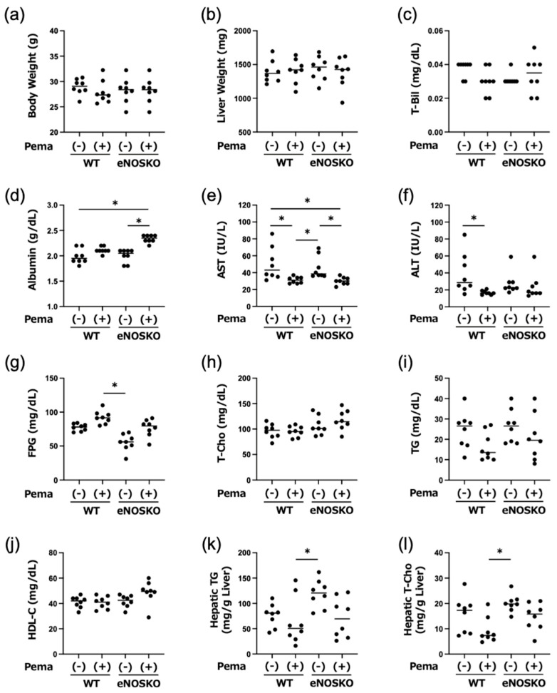
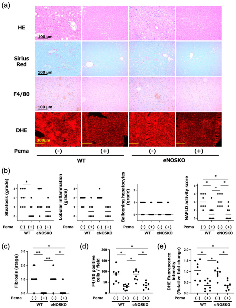
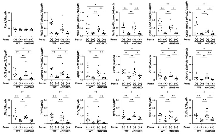
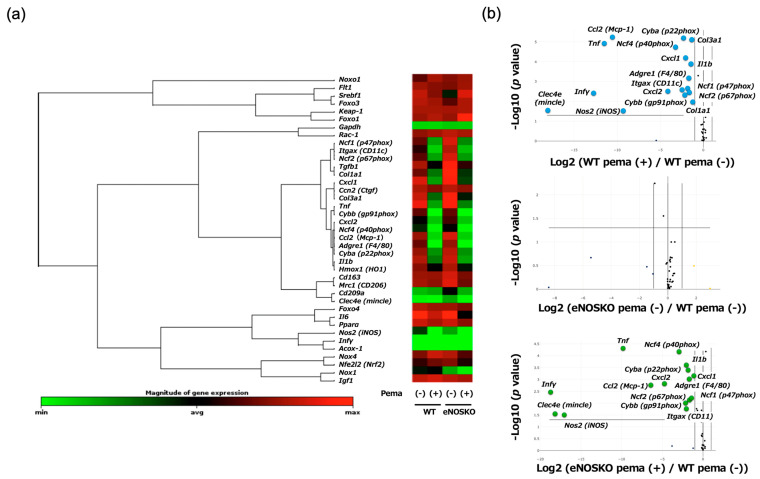
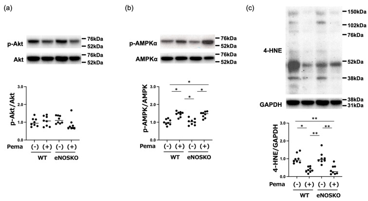
Endothelial dysfunction contributes to the progression of metabolic-dysfunction-associated steatotic liver disease (MASLD). Pemafibrate has been shown to ameliorate MASLD in basic and clinical studies, but it is unclear whether it is also effective in the status of endothelial dysfunction. An MASLD animal model was induced in male wild-type (WT) and endothelial nitric oxide synthase (eNOS)-deficient (eNOSKO) mice by feeding them a high-fat/cholesterol/cholate diet, and they were administered either a vehicle or pemafibrate at 0.17 mg/kg/day for 10 weeks. Although pemafibrate treatment did not change plasma lipid profiles in either WT or eNOSKO mice, pemafibrate reduced plasma AST levels in both WT and eNOSKO mice compared to the levels in the vehicle-treated mice. Histopathological analysis of the liver showed that MASLD was improved in the pemafibrate-treated groups in both WT and eNOSKO mice. Compared to vehicle treatment, pemafibrate treatment significantly reduced the expression levels of hepatic NADPH oxidase subunit genes, M1 macrophages, inflammatory-cytokine-related genes and profibrotic genes in both WT and eNOSKO mice, along with reduction in hepatic oxidative stress assessed by dihydroethidium staining and 4-hydroxynonenal protein levels. Thus, pemafibrate ameliorated MASLD with reduction in oxidative stress and inflammation even in vascular endothelial dysfunction.

Keywords: MASLD; NADPH oxidase; eNOS; endothelial dysfunction; oxidative stress; pemafibrate.


Conflict of interest statement

The authors declare no conflicts of interest.

Figures

Figure 1
Figure 1
Body weight (a), liver weight (b) and results of biochemical analyses (cl) of plasma samples and liver tissues in vehicle-treated MASLD mice and pemafibrate-treated MASLD mice with and without eNOS deficiency. Bars represent the mean values in each group (n = 8 mice per experimental group). * p < 0.05 is presented between the indicated groups. Kruskal–Wallis test with Dunn’s multiple comparisons test were used to assess statistical significance.
Figure 2
Figure 2
Hepatic histopathological examination, macrophage infiltration and superoxide detection in vehicle-treated MASLD mice and pemafibrate-treated MASLD mice with and without eNOS deficiency. (a) Representative findings of HE, Sirius red and F4/80 antibody staining and fluorescence with DHE in the liver of vehicle-treated MASLD mice and pemafibrate-treated MASLD mice with and without eNOS deficiency. (b) Histopathological assessment of the severity of MASLD in liver tissues. (c) Comparison of fibrosis staging in liver tissues. (d) Quantification of average F4/80-positive cell numbers counted in five independent visual fields in liver tissues. (e) Semi-quantitative analysis of DHE fluorescence intensities of liver tissues. Bars represent the mean values in each group (n = 8 mice per experimental group). * p < 0.05 and ** p < 0.01 are presented between the indicated groups. Kruskal–Wallis test with Dunn’s multiple comparisons test were used to assess statistical significance.
Figure 3
Figure 3
Hepatic gene expression levels of MASLD-associated factors in vehicle-treated MASLD mice and pemafibrate-treated MASLD mice with and without eNOS deficiency. Bars represent the mean values in each group (n = 8 mice per experimental group). * p < 0.05 and ** p < 0.01 are presented between the indicated groups. Kruskal–Wallis test with Dunn’s multiple comparisons test were used to assess statistical significance.
Figure 4
Figure 4
Profiling of hepatic gene expression in vehicle-treated MASLD mice and pemafibrate-treated MASLD mice with and without eNOS deficiency. (a) Group-wise integrated gene expression clustergram. (b) Volcano plots with reference to the vehicle-treated WT mouse group. n = 8 mice per experimental group.
Figure 5
Figure 5
Western blot analysis of MASLD-modulating factors in liver tissues of vehicle-treated MASLD mice and pemafibrate-treated MASLD mice with and without eNOS deficiency. (a) Representative blots of phosphorylated Akt and Akt, and ratios of phosphorylated Akt to Akt. (b) Representative blots of phosphorylated AMPKα and AMPKα, and ratios of phosphorylated AMPKα to AMPKα. (c) Representative blots of 4-HNE and GAPDH, and ratios of 4-HNE to GAPDH. Bars represent the mean values in each group (n = 8 mice per experimental group). * p < 0.05 and ** p < 0.01 are presented between the indicated groups. Kruskal–Wallis test with Dunn’s multiple comparisons test were used to assess statistical significance.

Similar articles

References

    1. Wong V.W., Ekstedt M., Wong G.L., Hagstrom H. Changing epidemiology, global trends and implications for outcomes of NAFLD. J. Hepatol. 2023;79:842–852. doi: 10.1016/j.jhep.2023.04.036. - DOI - PubMed
    1. Gambino R., Musso G., Cassader M. Redox balance in the pathogenesis of nonalcoholic fatty liver disease: Mechanisms and therapeutic opportunities. Antioxid. Redox Signal. 2011;15:1325–1365. doi: 10.1089/ars.2009.3058. - DOI - PubMed
    1. Younossi Z.M., Kalligeros M., Henry L. Epidemiology of metabolic dysfunction-associated steatotic liver disease. Clin. Mol. Hepatol. 2025;31:S32–S50. doi: 10.3350/cmh.2024.0431. - DOI - PMC - PubMed
    1. Adams L.A., Anstee Q.M., Tilg H., Targher G. Non-alcoholic fatty liver disease and its relationship with cardiovascular disease and other extrahepatic diseases. Gut. 2017;66:1138–1153. doi: 10.1136/gutjnl-2017-313884. - DOI - PubMed
    1. Angulo P., Kleiner D.E., Dam-Larsen S., Adams L.A., Bjornsson E.S., Charatcharoenwitthaya P., Mills P.R., Keach J.C., Lafferty H.D., Stahler A., et al. Liver Fibrosis, but No Other Histologic Features, Is Associated with Long-term Outcomes of Patients With Nonalcoholic Fatty Liver Disease. Gastroenterology. 2015;149:389–397.e10. doi: 10.1053/j.gastro.2015.04.043. - DOI - PMC - PubMed

LinkOut - more resources