. 2025 Jul 15;17(14):2349.
doi: 10.3390/cancers17142349.

Chimeric Element-Regulated MRI Reporter System for Mediation of Glioma Theranostics

Affiliations

Chimeric Element-Regulated MRI Reporter System for Mediation of Glioma Theranostics

Qian Hu et al. Cancers (Basel). .

Abstract

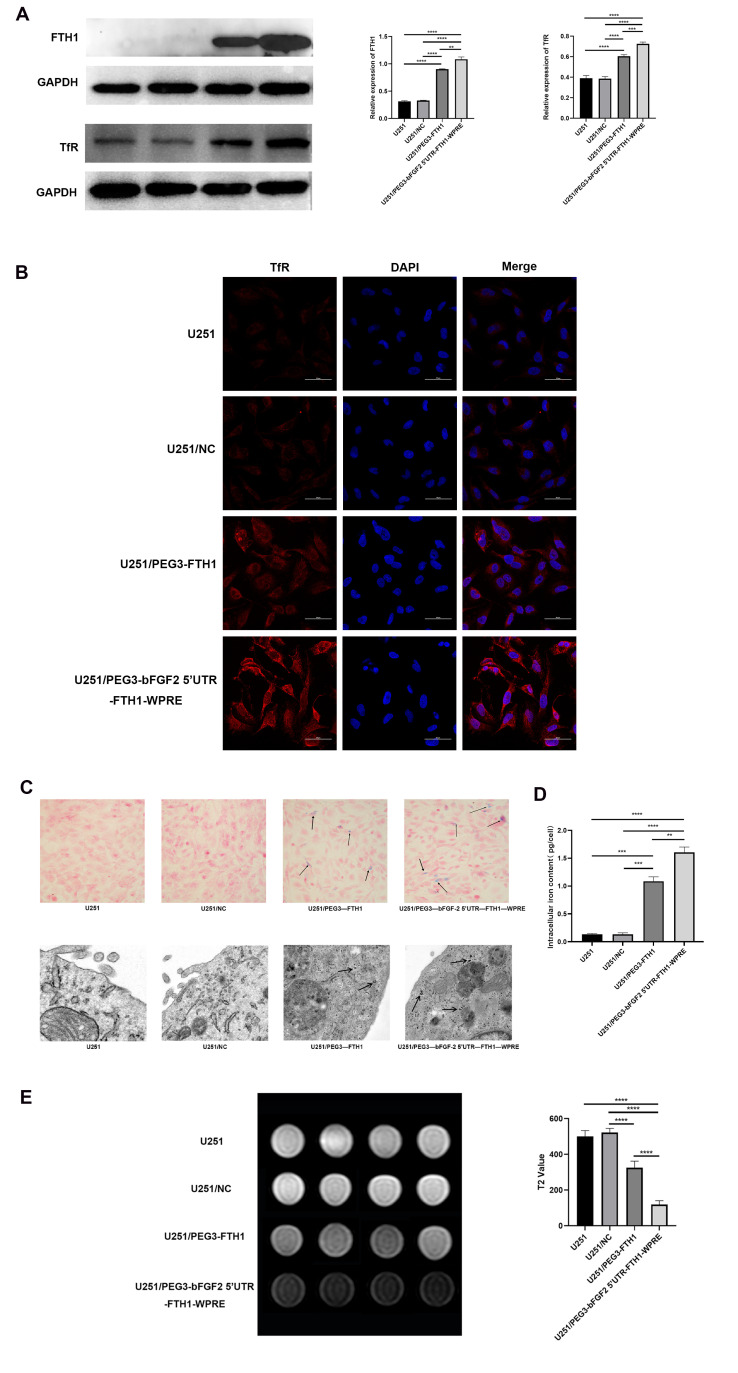
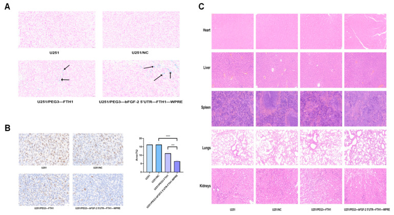
Background and Purpose: Glioblastoma remains a therapeutic challenge with a poor prognosis despite multimodal treatments. Reporter-based magnetic resonance imaging (MRI) offers a promising approach for tumor visualization, but its efficacy depends on sufficient reporter gene expression. This study aimed to develop a chimeric element-regulated ferritin heavy chain 1 (FTH1) reporter system to enhance MRI-based glioma detection while enabling targeted therapy via transferrin receptor (TfR)-mediated drug delivery. Methods: Using gene cloning techniques, we constructed a chimeric FTH1 expression system comprising tumor-specific PEG3 promoter (transcriptional control), bFGF-2 5'UTR (translational enhancement), and WPRE (mRNA stabilization). Lentiviral vectors delivered constructs to U251 glioblastoma cells and xenografts. FTH1/TfR expression was validated by Western blot and immunofluorescence. Iron accumulation was assessed via Prussian blue staining and TEM. MRI evaluated T2 signal changes. Transferrin-modified doxorubicin liposomes (Tf-LPD) were characterized for size and drug loading and tested for cellular uptake and cytotoxicity in vitro. In vivo therapeutic efficacy was assessed in nude mouse models through tumor volume measurement, MR imaging, and histopathology. Results: The chimeric system increased FTH1 expression significantly over PEG3-only controls (p < 0.01), with an increase of nearly 1.5-fold compared to the negative and blank groups and approximately a two-fold increase relative to the single promoter group, with corresponding TfR upregulation. Enhanced iron accumulation reduced T2 relaxation times significantly (p < 0.01), improving MR contrast. Tf-LPD (115 nm, 70% encapsulation) showed TfR-dependent uptake, inducing obvious apoptosis in high-TfR cells compared with that in controls. In vivo, Tf-LPD reduced tumor growth markedly in chimeric-system xenografts versus controls, with concurrent MR signal attenuation. Conclusions: The chimeric regulatory strategy overcomes limitations of single-element systems, demonstrating significant potential for integrated glioma theranostics. Its modular design may be adaptable to other reporter genes and malignancies.

Keywords: PEG3 promoter; chimeric element-regulated; reporter gene.


Conflict of interest statement

The authors declare that they have no known competing financial interests or personal relationships that could have appeared to influence the work reported in this paper.

Figures

Figure 1
Figure 1
FTH1 and TfR expression, iron trafficking effects, and MR imaging ((A): After lentiviral transfection, differences in FTH1 and TfR protein expression were observed among the U251 cell groups (**** p < 0.0001,*** p < 0.001,** p < 0.01; (B): Immunofluorescence results indicated significant variations in surface TfR expression across the U251 cell groups; (C): The Prussian blue staining assay revealed blue-stained granules (indicated by arrows) within the U251/PEG3-FTH1 and U251/PEG3-bFGF2 5′UTR-FTH1-WPRE cell groups, while TEM showed black iron granules (indicated by arrows) in these same groups; (D): The iron content in the U251/PEG3-FTH1 group and the U251/PEG3-bFGF2 5′UTR-FTH1-WPRE group was significantly higher than that in the U251 group and the U251/NC group (**** p < 0.0001). No significant difference was observed between the U251 group and the U251/NC group. (E): Magnetic resonance imaging T2-weighted images and histogram analysis of grayscale values demonstrated notable differences in signal intensity among the U251 cell groups).
Figure 2
Figure 2
Characterization and in vitro uptake of Tf-LP and Tf-LPD ((A,B): TEM revealed that both Tf-LP and Tf-LPD exhibit spherical morphology and uniform size as nanomaterials; (C,D): Particle size analysis indicated that the diameter of Tf-LPD is (114.94 ± 3.17) nm, while Tf-LP has a diameter of (112.33 ± 6.53) nm; (E): Immunofluorescence results demonstrated that the uptake of Tf-LP by U251 cells increased over time, with the U251/PEG3-bFGF2 5′UTR-FTH1-WPRE group showing the highest uptake, Scale bar: 50 μm).
Figure 3
Figure 3
Cytotoxicity of Tf-LP and Tf-LPD ((A):The CCK8 assay results indicate that Tf-LP exhibits minimal cytotoxicity across all U251 cell groups; (B): The results of the CCK8 assay indicate that the survival rate of U251 cells in all groups decreased with increasing concentrations of Tf-LPD, with the U251/PEG3-bFGF2 5′UTR-FTH1-WPRE group exhibiting the lowest cell survival rate; (C): The results of the flow cytometry experiments indicated that the apoptosis rates for the four groups of cells—U251, U251/NC, U251/PEG3-FTH1, and U251/PEG3-bFGF2 5′UTR-FTH1-WPRE—were 3.15%, 4.62%, 41%, and 72.5%).
Figure 4
Figure 4
Fluorescent distribution maps of lentivirus and Tf-LPD in mice, growth curve of tumors ((A): Significant fluorescence distribution was observed in the tumor sites of mice from the three groups following in vivo transfection with lentiviral vectors; (B): After entering the nude mice via the tail vein, Tf-LPD primarily accumulates in the liver at 6 h, and at 24 h, it is predominantly found in the tumor. The fluorescence signals in the U251/PEG3-FTH1 and U251/PEG3-bFGF2 5′UTR-FTH1-WPRE groups are significantly stronger than those in the U251 and U251/-NC groups, with nearly complete metabolism observed by 48 h; (C): On day 21, the tumors from each group of mice were dissected, revealing that the U251/PEG3-bFGF2 5′UTR-FTH1-WPRE group exhibited the smallest tumor size; (D): By systematically measuring and recording tumor growth in various groups of mice, we generated growth curves. The results indicate that the tumor growth rates in the U251 and U251/-NC groups showed no significant differences and were faster than those in the other two groups, while the U251/PEG3-bFGF2 5′UTR-FTH1-WPRE group exhibited the slowest tumor growth rate).
Figure 5
Figure 5
(A): Axial T2-weighted imaging of tumors in each group at various time points; (B): Analysis of T2 values for tumors in each group at different time intervals (a, b, c, and d represent the T2 signal intensity measurements obtained before and at 3, 7, and 14 days after intravenous administration of drug-loaded liposomes in tumor-bearing mice, respectively). Analysis of the T2WI scans and T2 grayscale values revealed that the U251 and U251/NC groups exhibited no significant differences in tumor signal intensity, both showing slightly elevated signals. In contrast, the U251/PEG3-FTH1 group displayed a lower tumor signal compared to the first two groups, while the U251/PEG3-bFGF2 5′UTR-FTH1-WPRE group demonstrated a more pronounced reduction in tumor signal intensity) (*** p < 0.001,** p < 0.01,* p <0.05).
Figure 6
Figure 6
Immunohistochemistry, Prussian blue staining of tumor tissue, and HE staining of major organs ((A): Both the U251/PEG3-FTH1 group and the U251/PEG3-bFGF2 5′ UTR-FTH1-WPRE group exhibited blue-stained granules (indicated by arrows) within the tumor tissue cells, Scale bar: 20 μm; (B): The analysis of the proportion of Ki-67 immunohistochemically positive cells in tumor tissues revealed that the positive cell area percentages for the four groups were 16.34 ± 0.9, 16.29 ± 1.17, 11.15 ± 1.47, and 6.47 ± 0.56, (*** p < 0.001,**** p < 0.0001), Scale bar: 20 μm; (C): HE staining results indicate that the primary immune organ cell structures in all four groups of mice showed no significant damage, Scale bar: 50 μm).

References

    1. Ostrom Q.T., Price M., Neff C., Cioffi G., Waite K.A., Kruchko C., Barnholtz-Sloan J.S. CBTRUS Statistical Report: Primary Brain and Other Central Nervous System Tumors Diagnosed in the United States in 2016–2020. Neuro Oncol. 2023;25((12 Suppl. 2)):iv1–iv99. doi: 10.1093/neuonc/noad149. - DOI - PMC - PubMed
    1. Clarke J., Penas C., Pastori C., Komotar R.J., Bregy A., Shah A.H., Wahlestedt C., Ayad N.G. Epigenetic pathways and glioblastoma treatment. Epigenetics. 2013;8:785–795. doi: 10.4161/epi.25440. - DOI - PMC - PubMed
    1. Szopa W., Burley T.A., Kramer-Marek G., Kaspera W. Diagnostic and Therapeutic Biomarkers in Glioblastoma: Current Status and Future Perspectives. Biomed. Res. Int. 2017;2017:8013575. doi: 10.1155/2017/8013575. - DOI - PMC - PubMed
    1. Shalaby N., Dubois V.P., Ronald J. Molecular imaging of cellular immunotherapies in experimental and therapeutic settings. Cancer Immunol. Immunother. 2022;71:1281–1294. doi: 10.1007/s00262-021-03073-5. - DOI - PMC - PubMed
    1. Haris M., Yadav S.K., Rizwan A., Singh A., Wang E., Hariharan H., Reddy R., Marincola F.M. Molecular magnetic resonance imaging in cancer. J. Transl. Med. 2015;13:313. doi: 10.1186/s12967-015-0659-x. - DOI - PMC - PubMed

LinkOut - more resources