. 2025 Jul 7;14(7):826.
doi: 10.3390/biology14070826.

Unraveling the Role of Spicules in Shaping Sponge Body Structure: Evidence from the Early Cambrian Shuijingtuo Formation

Affiliations

Unraveling the Role of Spicules in Shaping Sponge Body Structure: Evidence from the Early Cambrian Shuijingtuo Formation

Xinyi Ren et al. Biology (Basel). .

Abstract

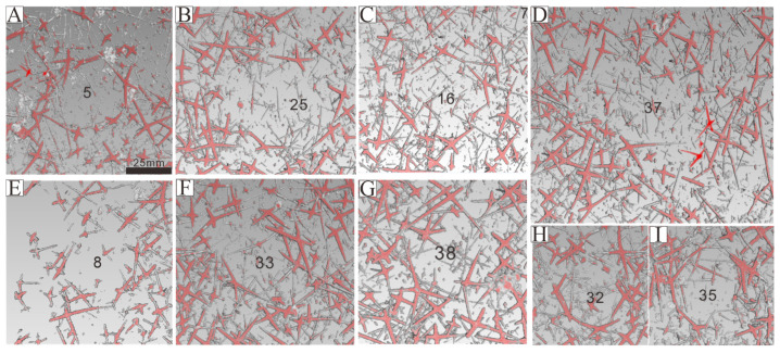
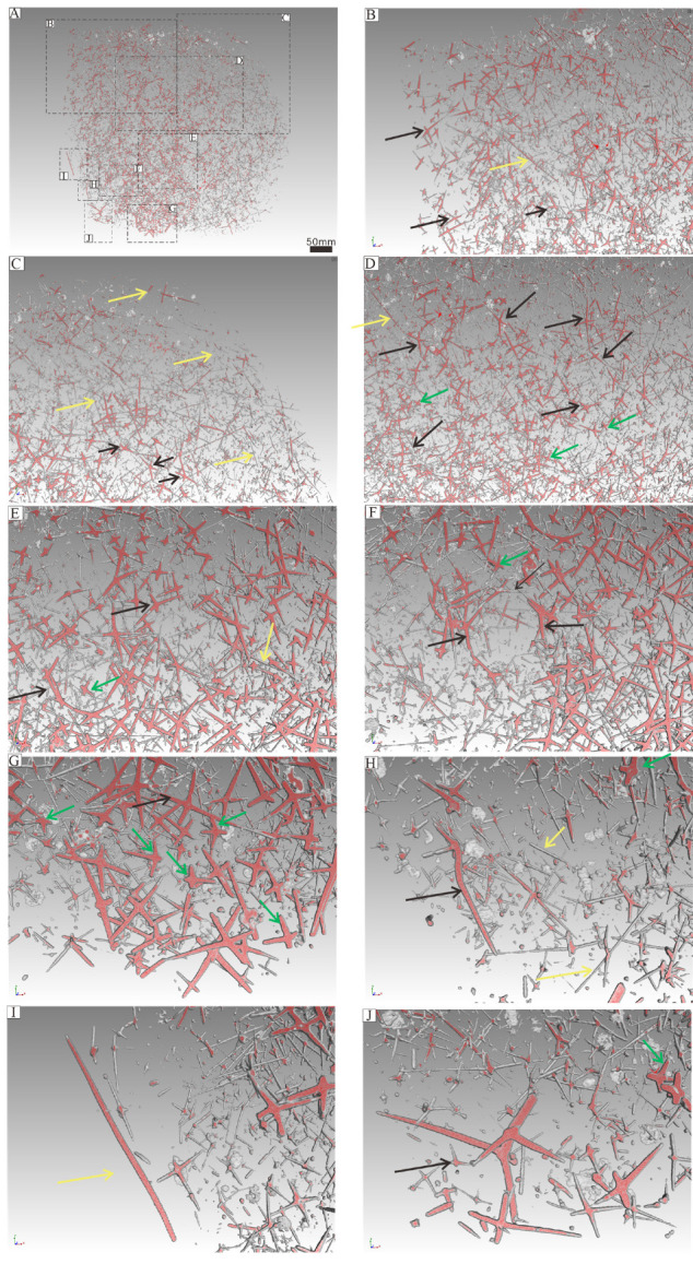
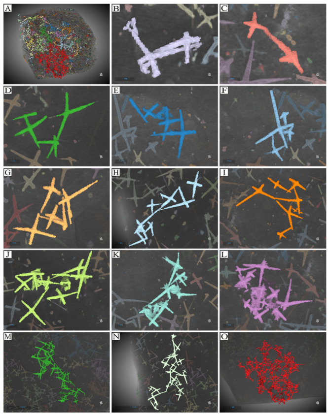
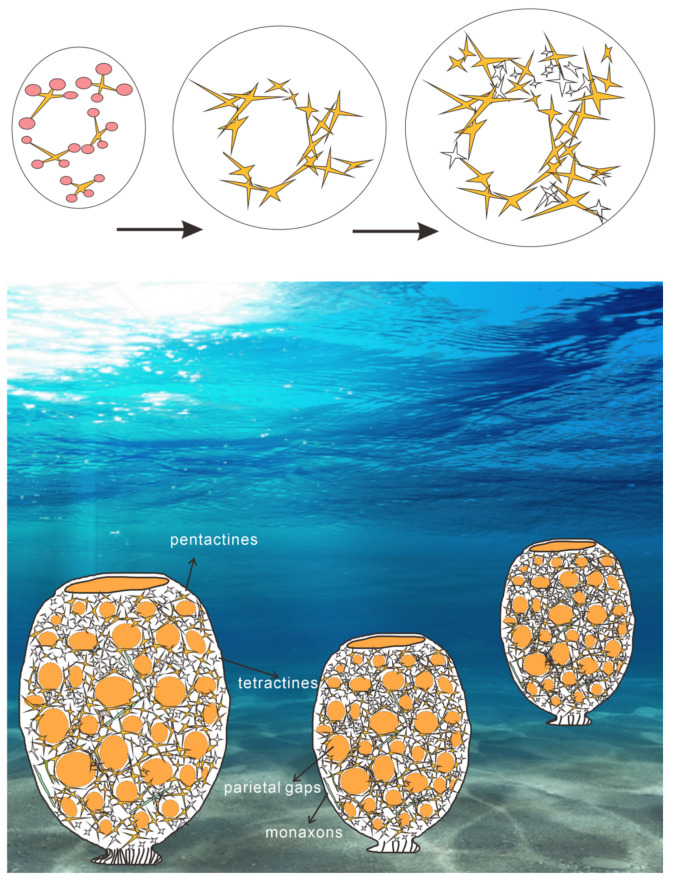
In most cases, sponge fossils are preserved as isolated spicules, with complete sponge body fossils largely confined to Konservat-Lagerstätten. Although the classification and diversity of sponges and their isolated spicules have been extensively studied, no systematic attempts have been made to define the relationship between fossil spicules and the sponge body plan. By utilizing relatively well-preserved sponge fossils from the black shales of the Shuijingtuo Formation (South China) in conjunction with isolated spicules from the same locality, we assess spicule morphology to identify the potential functional roles of spicules and chart their arrangement within the sponge body. The elemental distribution and three-dimensional morphology of the examined sponge body fossil (likely a hexactinelid) are assessed using both micro-XRF and micro-CT. Tetractine, stauractine and pentactine spicules are the most abundant spicule types, both in the body fossil and in acid residues, with an additional spicule type (monaxons) also present. The larger pentactine spicules (five-ray spicules) frame the structure, whereas the smaller tetractines and stauractines (four-ray spicules), along with smaller pentactines, are arranged along the branches of the larger spicules. Based on the arrangement of the different spicules, it is proposed that each of the spicule types represents a discrete functional form: monaxons support the overall sponge body plan, pentactines construct the framework of the parietal gaps, and the smaller pentactines or tetractines stabilize the framework of the parietal gaps. These results provide a new understanding of sponge morphology, spicule function and the relationship between isolated fossil spicules and associated sponge body fossils.

Keywords: Cambrian; micro-CT; micro-XRF; spicules; sponge.


Conflict of interest statement

The authors declare no conflicts of interest.

Figures

Figure A1
Figure A1
Morphology and spicule arrangement for the 42 parietal gaps (all images are at the same scale) based on micro-CT results.
Figure 1
Figure 1
Regional geological map of China, map of sampling locations, partial geological map of Hubei Province, and stratigraphic column of the Shuijingtuo Formation in Aijiahe, Yichang. (A) Regional geological map of China, with the sampling area for this study located in Zone II. (B) Geological map of Yichang and its surrounding regions, with yellow stars highlighting the sampling points within the Cambrian strata (light yellow color). (C) Sampling site in Aijiahe, Yichang City, Hubei Province, indicated by the yellow star. (D) Stratigraphic column of the Shuijingtuo Formation for sample site near Aijiahe village.
Figure 2
Figure 2
Isolated sponge spicules (AR) (the scale bar represents 200 μm). Spicule (A) is rodlike and is incomplete (1 rays); Spicule (D) has 3 ends (3 rays); Spicules (B,C,EJ) have 4 branches in different directions (4 rays); Spicules (KO) have 5 branches (5 rays); Spicules (PR) have 6 branches (6 rays).
Figure 3
Figure 3
The possible spicule types recovered from acid residues.
Figure 4
Figure 4
Proportions of each spicule form of isolated spicules presented as a column chart (A) and a pie chart (B). The 4-ray and 5-ray spicules represent the majority of spicules, with 5-ray spicules making up more than 50% of all specimens.
Figure 5
Figure 5
XRF elemental distribution of sponge spicules in the Shuijingtuo Formation, from Aijiahe village, Hubei province (sample number: Figures (AO) are AJH-D6-213; red = iron, blue = silicon, russet = sulfur, green = phosphate, yellow = calcium; and Figures (PU) are AJH-D5-184; red = iron, blue = silicon, sky blue = sulfur, green = phosphate, yellow = calcium). (AC) show fossil morphology in situ. (DG,L,M,PS) show simple elemental information for the sponge fossils, and (HK,N,O,T,U) show the distribution of multiple elements simultaneously for the same fossils. The sponge spicules consist of Ca, S, and Fe, with S and Fe often overlapping. The matrix consists of Si in all samples.
Figure 6
Figure 6
Iron (red color) and calcium (blue color) elements are compared based on micro-XRF elemental mapping. (A) illustrates the fossil divided into a 1 cm2 grid (1 cm width × 1 cm length). (B) depicts the number of spicules within each cell of the grid, where the horizontal axis represents each grid cell in order from left to right and top to bottom, and the vertical axis indicates the number of spicules in each corresponding grid cell. The majority of cells fall within the range of 40 to 80 spicules. (C) shows both the number of parietal gaps and their respective compositions, in terms of spicule count. (D) indicates that most parietal gaps are composed of 8, 10, 15, 20, or 25 spicules, while a few incurrent pores consist of only 6, 7, or 17 spicules.
Figure 7
Figure 7
Micro-CT results. Parietal gaps (indicated by the numbers in black) are clearly surrounded by spicules (AI). Micro-CT results indicating spicule counts for each parietal gap ranging from 5 to 29 (AI, Appendix C), with the majority consisting of 10, 12, 15, or 16 spicules (Appendix C). All images are at the same scale.
Figure 8
Figure 8
Micro-CT results showing spicule position and orientation for different spicule types associated with parietal gaps. Yellow, black, and green arrows are used to denote different types of spicules. Yellow arrows indicate monaxons, green arrows represent smaller five-ray spicules and black arrows signify larger three-/four-/five-ray spicules (Appendix A). (BJ) represent each of the labeled areas indicated in (A).
Figure 9
Figure 9
Micro-CT results showing spicule position and orientation for different spicule types associated with parietal gaps. (AJ) represent the same parts of the specimen depicted in Figure 8 but are rotated by 45 degrees to show the dominant orientations for each spicule in the third dimension. Yellow, black, and green arrows denote different types of spicules, with yellow arrows indicating monaxons, green arrows representing smaller five-ray spicules, and black arrows signifying larger three-/four-/five-ray spicules (Appendix A).
Figure 10
Figure 10
(A) Reconstructed Micro-CT data for the entire studied specimen. (BO) Spicule arrangement for multiple spicule morphologies. Individual spicules are demarcated using false colors. The micro-CT reconstruction presented in (A) provides a preliminary delineation of the various types of spicules (see Appendix A), which are distinguished by different colors. The prominently colored regions are primarily composed of Fe. (B,C) depict a nested arrangement formed by two spicules. (DF) illustrate a structure composed of four spicules. (GK) show an arrangement of six or seven spicules. (LN) show structures composed of multiple larger and smaller spicules. (O) illustrates multiple parietal gaps and the associated pattern of spicules. In (D,FK), the meshwork of parietal gaps is composed of large spicules forming angles greater than 90 degrees. Although the angles are not uniform, the obtuse angles generally point toward each parietal gap, while the acute angles direct outward toward the tangential surfaces of the meshes. The construction modes can be categorized into three types: point–point contact, point–surface contact, and surface–surface contact. (H,I) show the branches of adjacent spicules converge at a certain point, representing point–point contact. In (K), one spicule’s branch inserts into the acute angle of an adjacent spicule, indicating point–surface contact. In (B,J), intersecting surfaces are formed by respective angles, signifying face–surface contact (Appendix A).
Figure 11
Figure 11
Principal Components Analysis for the three extant sponge clades: Calcarea (red), Hexactinellida (green) and Demospongiae (blue). Demospongiae and Hexactinellida are somewhat differentiated but Calcarea completely overlaps with Demospongiae and Hexactinellida.
Figure 12
Figure 12
Results of Origin 8.6 analysis for extant sponge clades and our fossil sample. The horizontal axis represents the branch number for a single spicule from the relevant sponge clade. One the vertical axis, 0 represents spicules have more than one developmental center, and 1 means that spicules that have a single origin site.
Figure 13
Figure 13
Reconstruction of Hexactinellida taxon from the Shuijingtuo Formation, eastern Three Gorges, Hubei Province (Reference from [13,24,41,45,48,51,57,58,59,60,61,62,63,64,65,66,67]. Copyright information for background photo is provided in Appendix I.

References

    1. Walter M.R. The Biology of Reefs and Reef Organisms. Chicago University Press; Chicago, IL, USA: 2013. 410p. - DOI
    1. Bernard M.D., Sally L., Claire L. Sponge development and antiquity of animal pattern formation. Integr. Comp. Biol. 2005;45:335–341. doi: 10.1093/icb/45.2.335. - DOI - PubMed
    1. Yang J.Y., Ma H.D., Chen A.L., Hou X.G. A New Leptomitid Sponge from the Early Cambrian Chengjiang Biota. Geol. J. China Univ. 2019;25:480. doi: 10.16108/j.issn1006-7493.2018117. (In Chinese with English Abstract) - DOI
    1. Li L.X., Yan G.Z., Wei X., Gong F.Y., Oliver L., Wu R.C. Late Ordovician sponge spicules from the Yangtze Platform, South China: Biostratigraphical and palaeobiogeographical significance. J. Asian Earth Sci. 2025;277:106380. doi: 10.1016/j.jseaes.2024.106380. - DOI
    1. Zhao X., Li G.X. Early Cambrian Sponge Spicule Fossils from Zhenba County, Southern Shaanxi Province. Acta Micropalaeontologica Sin. 2006;23:281–294. (In Chinese with English Abstract)

LinkOut - more resources